4.1 Types of Tissues
Learning Objectives
Identify the main tissue types and discuss their roles in the human body.
By the end of this section, you will be able to:
- Identify the four primary tissue types and discuss the structure and function of each
- Describe the embryonic origin of tissue
- Identify the various types of tissue membranes and the unique qualities of each
The term tissue is used to describe a group of cells that are similar in structure and perform a specific function. Histology is the the field of study that involves the microscopic examination of tissue appearance, organization, and function.
Tissues are organized into four broad categories based on structural and functional similarities. These categories are epithelial, connective, muscle, and nervous. The primary tissue types work together to contribute to the overall health and maintenance of the human body. Thus, any disruption in the structure of a tissue can lead to injury or disease.
The Four Primary Tissue Types
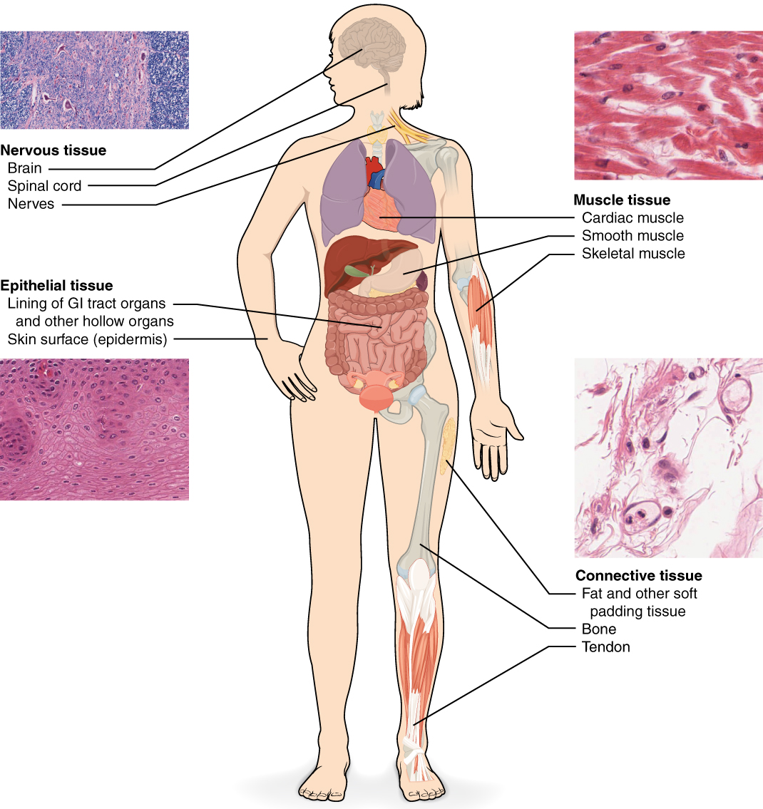
Epithelial tissue refers to groups of cells that cover the exterior surfaces of the body, line internal cavities and passageways, and form certain glands. Connective tissue, as its name implies, binds the cells and organs of the body together. Muscle tissue contracts forcefully when excited, providing movement. Nervous tissue is also excitable, allowing for the generation and propagation of electrochemical signals in the form of nerve impulses that communicate between different regions of the body (Figure 4.1.1).
An understanding of the various primary tissue types present in the human body is essential for understanding the structure and function of organs which are composed of two or more primary tissue types. This chapter will focus on examining epithelial and connective tissues. Muscle and nervous tissue will be discussed in detail in future chapters.
Embryonic Origin of Tissues
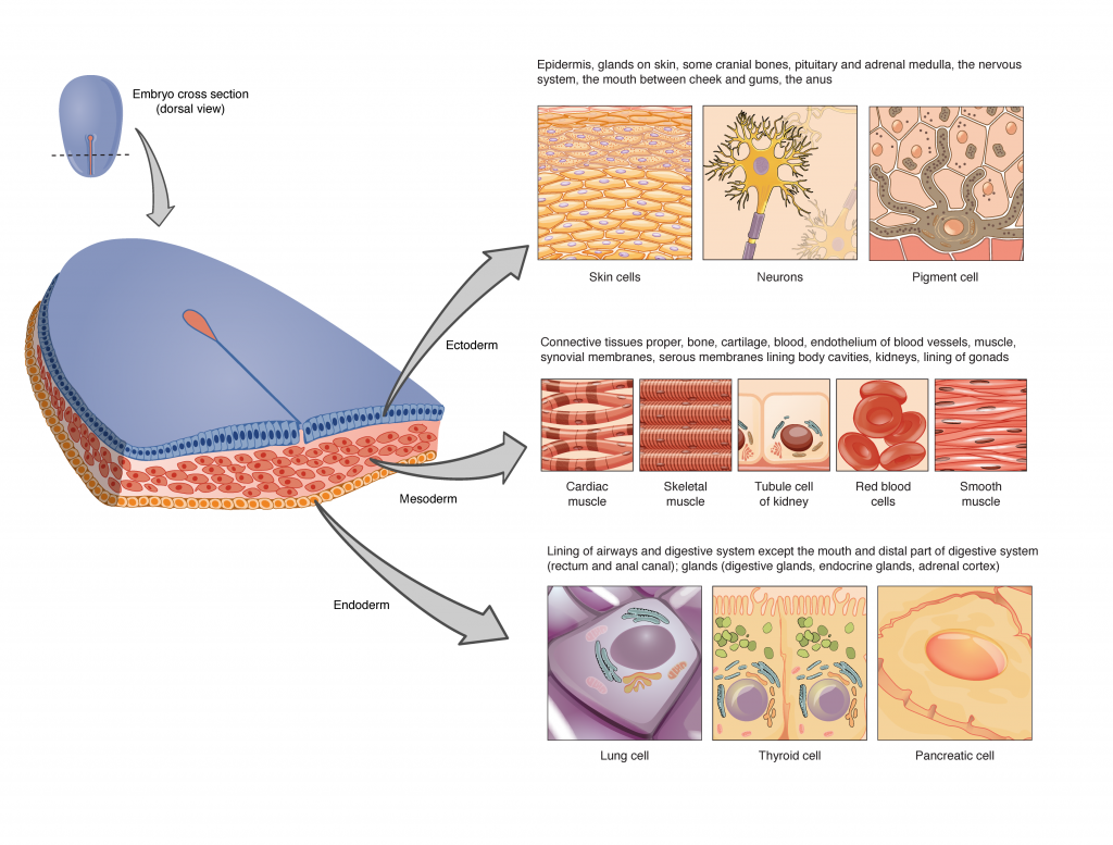
The cells composing a tissue share a common embryonic origin. The zygote, or fertilized egg, is a single cell formed by the fusion of an egg and sperm cell. After fertilization, the zygote gives rise many cells to form the embryo. The first embryonic cells generated have the ability to differentiate into any type of cell in the body and, as such, are called omnipotent, meaning each has the capacity to divide, differentiate, and develop into a new organism. As cell proliferation progresses, three major cell lines are established within the embryo. Each of these lines of embryonic cells forms the distinct germ layers from which all the tissues and organs of the human body eventually form. Each germ layer is identified by its relative position: ectoderm (ecto- = “outer”), mesoderm (meso- = “middle”), and endoderm (endo- = “inner”). Figure 4.1.2 shows the types of tissues and organs associated with each of the three germ layers. Note that epithelial tissue originates in all three layers, whereas nervous tissue derives primarily from the ectoderm and muscle tissue derives from the mesoderm.
External Website

View this slideshow to learn more about stem cells. How do somatic stem cells differ from embryonic stem cells?
Tissue Membranes
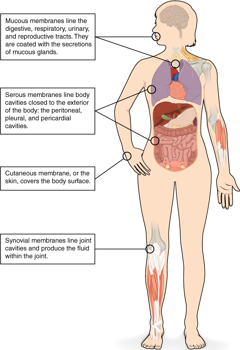
A tissue membrane is a thin layer or sheet of cells that either covers the outside of the body (e.g., skin), lines an internal body cavity (e.g., peritoneal cavity), lines a vessel (e.g., blood vessel), or lines a movable joint cavity (e.g., synovial joint). Two basic types of tissue membranes are recognized based on the primary tissue type composing each: connective tissue membranes and epithelial membranes (Figure 4.1.3).
Connective Tissue Membranes
A connective tissue membrane is built entirely of connective tissue. This type of membrane may be found encapsulating an organ, such as the kidney, or lining the cavity of a freely movable joint (e.g., shoulder). When lining a joint, this membrane is referred to as a synovial membrane. Cells in the inner layer of the synovial membrane release synovial fluid, a natural lubricant that enables the bones of a joint to move freely against one another with reduced friction.
Epithelial Membranes
An epithelial membrane is composed of an epithelial layer attached to a layer of connective tissue. A mucous membrane, sometimes called a mucosa, lines a body cavity or hollow passageway that is open to the external environment. This type of membrane can be found lining portions of the digestive, respiratory, excretory, and reproductive tracts. Mucus, produced by uniglandular cells and glandular tissue, coats the epithelial layer. The underlying connective tissue, called the lamina propria (literally “own layer”), helps support the epithelial layer.
A serous membrane lines the cavities of the body that do not open to the external environment. Serous fluid secreted by the cells of the epithelium lubricates the membrane and reduces abrasion and friction between organs. Serous membranes are identified according to location. Three serous membranes are found lining the thoracic cavity; two membranes that cover the lungs (pleura) and one membrane that covers the heart (pericardium). A fourth serous membrane, the peritoneum, lines the peritoneal cavity, covering the abdominal organs and forming double sheets of mesenteries that suspend many of the digestive organs.
A cutaneous membrane is a multi-layered membrane composed of epithelial and connective tissues. The apical surface of this membrane exposed to the external environment and is covered with dead, keratinized cells that help protect the body from desiccation and pathogens. The skin is an example of a cutaneous membrane.
Chapter Review
Aggregations of cells in the human body be classified into four types of tissues: epithelial, connective, muscle, and nervous. Epithelial tissues act as coverings, controlling the movement of materials across their surface. Connective tissue binds the various parts of the body together, providing support and protection. Muscle tissue allows the body to move and nervous tissues functions in communication.
All cells and tissues in the body derive from three germ layers: the ectoderm, mesoderm, and endoderm.
Membranes are layers of connective and epithelial tissues that line the external environment and internal body cavities of the body. Synovial membranes are connective tissue membranes that protect and line the freely-movable joints. Epithelial membranes are composed of both epithelial tissue and connective tissue. These membranes are found lining the external body surface (cutaneous membranes and mucous membranes) or lining the internal body cavities (serous membranes).
Interactive Link Questions
View this slideshow to learn more about stem cells. How do somatic stem cells differ from embryonic stem cells?
Most somatic stem cells give rise to only a few cell types.
Review Questions
Critical Thinking Questions
Identify the four types of tissue in the body, and describe the major functions of each tissue.
The four types of tissues in the body are epithelial, connective, muscle, and nervous. Epithelial tissue is made of layers of cells that cover the surfaces of the body that come into contact with the exterior world, line internal cavities, and form glands. Connective tissue binds the cells and organs of the body together and performs many functions, especially in the protection, support, and integration of the body. Muscle tissue, which responds to stimulation and contracts to provide movement, is divided into three major types: skeletal (voluntary) muscles, smooth muscles, and the cardiac muscle in the heart. Nervous tissue allows the body to receive signals and transmit information as electric impulses from one region of the body to another.
The zygote is described as omnipotent because it ultimately gives rise to all the cells in your body including the highly specialized cells of your nervous system. Describe this transition, discussing the steps and processes that lead to these specialized cells.
The zygote divides into many cells. As these cells become specialized, they lose their ability to differentiate into all tissues. At first they form the three primary germ layers. Following the cells of the ectodermal germ layer, they too become more restricted in what they can form. Ultimately, some of these ectodermal cells become further restricted and differentiate in to nerve cells.
What happens when a terminally differentiated cell reverts to a less differentiated state?
What is the function of synovial membranes?
Synovial membranes are a type of connective tissue membrane that supports mobility in joints. The membrane lines the joint cavity and contains fibroblasts that produce hyaluronan, which leads to the production of synovial fluid, a natural lubricant that enables the bones of a joint to move freely against one another.
This work, Anatomy & Physiology, is adapted from Anatomy & Physiology by OpenStax, licensed under CC BY. This edition, with revised content and artwork, is licensed under CC BY-SA except where otherwise noted.
Images, from Anatomy & Physiology by OpenStax, are licensed under CC BY except where otherwise noted.
Access the original for free at https://openstax.org/books/anatomy-and-physiology/pages/1-introduction.
