13.1 Sensory Receptors
Learning Objectives
By the end of this section, you will be able to:
- Describe different types of sensory receptors
A major role of sensory receptors is to help us learn about the environment around us, or about the state of our internal environment. Different types of stimuli from varying sources are received and changed into the electrochemical signals of the nervous system. This process is called sensory transduction. This occurs when a stimulus is detected by a receptor which generates a graded potential in a sensory neuron. If strong enough, the graded potential causes the sensory neuron to produce an action potential that is relayed into the central nervous system (CNS), where it is integrated with other sensory information—and sometimes higher cognitive functions—to become a conscious perception of that stimulus. The central integration may then lead to a motor response.
Describing sensory function with the term sensation or perception is a deliberate distinction. Sensation is the activation of sensory receptors at the level of the stimulus. Perception is the central processing of sensory stimuli into a meaningful pattern involving awareness. Perception is dependent on sensation, but not all sensations are perceived. Receptors are the structures (and sometimes whole cells) that detect sensations. A receptor or receptor cell is changed directly by a stimulus. A transmembrane protein receptor is a protein in the cell membrane that mediates a physiological change in a neuron, most often through the opening of ion channels or changes in the cell signaling processes. Some transmembrane receptors are activated by chemicals called ligands. For example, a molecule in food can serve as a ligand for taste receptors. Other transmembrane proteins, which are not accurately called receptors, are sensitive to mechanical or thermal changes. Physical changes in these proteins increase ion flow across the membrane, and can generate a graded potential in the sensory neurons.
Sensory Receptors
Stimuli in the environment activate specialized receptors or receptor cells in the peripheral nervous system. Different types of stimuli are sensed by different types of receptors. Receptor cells can be classified into types on the basis of three different criteria: cell type, position, and function. Receptors can be classified structurally on the basis of cell type and their position in relation to stimuli they sense. They can also be classified functionally on the basis of the transduction of stimuli, or how the mechanical stimulus, light, or chemical changed the cell membrane potential.
Structural Receptor Types
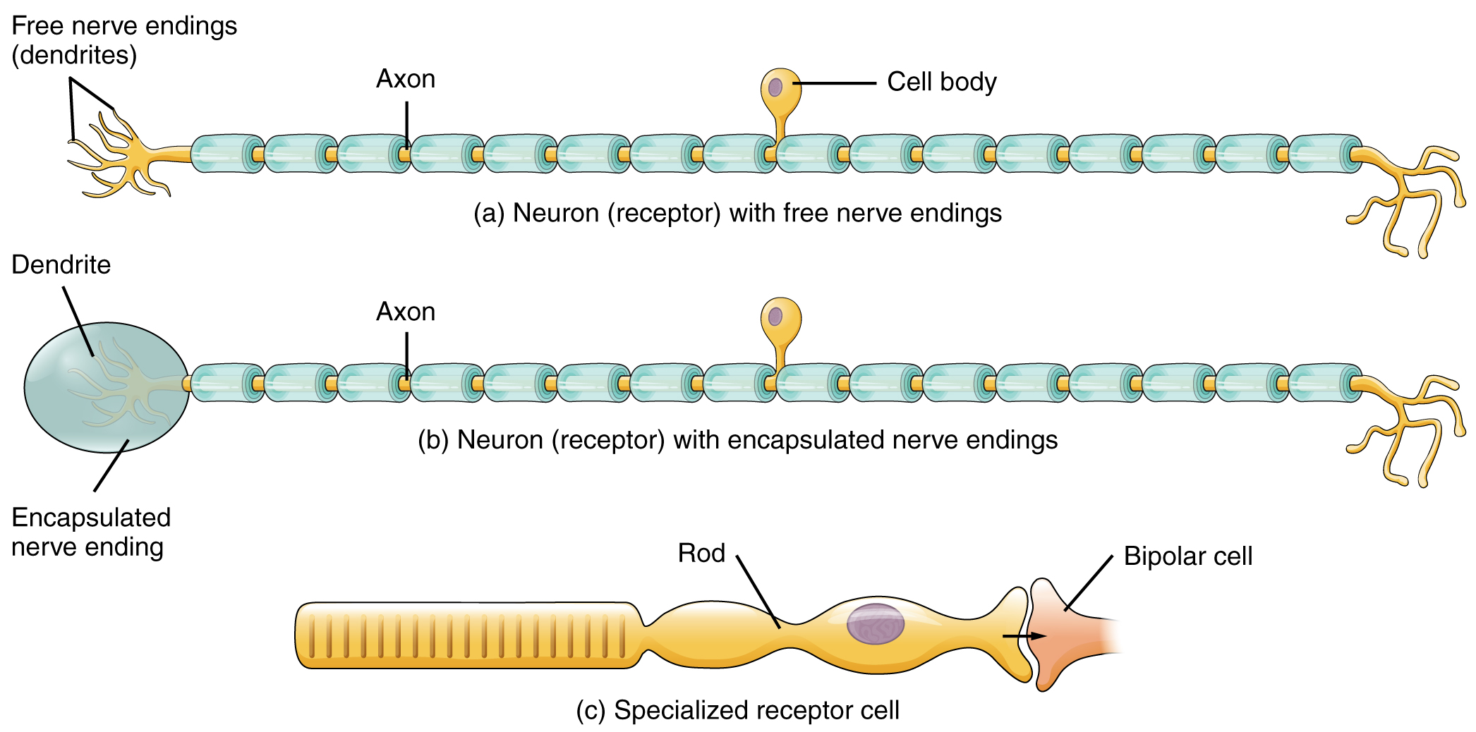
The cells that interpret information about the environment can be either (1) a neuron that has a free nerve ending (dendrites) embedded in tissue that would receive a sensation; (2) a neuron that has an encapsulated ending in which the dendrites are encapsulated in connective tissue that enhances their sensitivity; or (3) a specialized receptor cell, which has distinct structural components that interpret a specific type of stimulus (Figure 13.1.1). The pain and temperature receptors in the dermis of the skin are examples of neurons that have free nerve endings. Also located in the dermis of the skin are lamellated and tactile corpuscles, neurons with encapsulated nerve endings that respond to pressure and touch. The cells in the retina that respond to light stimuli are an example of a specialized receptor cell, a photoreceptor.
Graded potentials in free and encapsulated nerve endings are called generator potentials. When strong enough to reach threshold they can directly trigger an action potential along the axon of the sensory neuron. Action potentials triggered by receptor cells, however, are indirect. Graded potentials in receptor cells are called receptor potentials. These graded potentials cause neurotransmitter to be released onto a sensory neuron causing a graded post-synaptic potential. If this graded post-synaptic potential is strong enough to reach threshold it will trigger an action potential along the axon of the sensory neuron.
Another way that receptors can be classified is based on their location relative to the stimuli. An exteroceptor is a receptor that is located near a stimulus in the external environment, such as the somatosensory receptors that are located in the skin. An interoceptor is one that detects stimuli from internal organs and tissues, such as the receptors that sense the increase in blood pressure in the aorta or carotid sinus. Finally, a proprioceptor is a receptor located near a moving part of the body, such as a muscle or joint capsule, that interprets the positions of the tissues as they move.
Functional Receptor Types
A third classification of receptors is by how the receptor transduces stimuli into membrane potential changes. Stimuli are of three general types. Some stimuli are ions and macromolecules that affect transmembrane receptor proteins by binding or by directly diffusing across the cell membrane. Some stimuli are physical variations in the environment that affect receptor cell membrane potentials. Other stimuli include the electromagnetic radiation from visible light. For humans, the only electromagnetic energy that is perceived by our eyes is visible light. Some other organisms have receptors that humans lack, such as the heat sensors of snakes, the ultraviolet light sensors of bees, or magnetic receptors in migratory birds.
Receptor cells can be further categorized on the basis of the type of stimuli they transduce. Chemical stimuli can be detected by a chemoreceptors that detect chemical stimuli, such as a chemicals that lead to the sense of smell. Osmoreceptors respond to solute concentrations of body fluids. Pain is primarily a chemical and sometimes mechanical sense that interprets the presence of chemicals from tissue damage, or intense mechanical stimuli, through a nociceptor. Physical stimuli, such as pressure and vibration, as well as the sensation of sound and body position (balance), are interpreted through a mechanoreceptor. Another physical stimulus that has its own type of receptor is temperature, which is sensed through a thermoreceptor that is either sensitive to temperatures above (heat) or below (cold) normal body temperature.
Sensory Modalities
Ask anyone what the senses are, and they are likely to list the five major senses—taste, smell, touch, hearing, and sight. However, these are not all of the senses. The most obvious omission from this list is balance. Also, what is referred to simply as touch can be further subdivided into pressure, vibration, stretch, and hair-follicle position, on the basis of the type of mechanoreceptors that perceive these touch sensations. Other overlooked senses include temperature perception by thermoreceptors and pain perception by nociceptors.
Within the realm of physiology, senses can be classified as either general or special. A general sense is one that is distributed throughout the body and has receptor cells within the structures of other organs. Mechanoreceptors in the skin, muscles, or the walls of blood vessels are examples of this type. General senses often contribute to the sense of touch, as described above, or to proprioception (body position) and kinesthesia (body movement), or to a visceral sense, which is most important to autonomic functions. A special sense (discussed in Chapter 15) is one that has a specific organ devoted to it, namely the eye, inner ear, tongue, or nose.
Each of the senses is referred to as a sensory modality. Modality refers to the way that information is encoded into a perception. The main sensory modalities can be described on the basis of how each stimulus is transduced and perceived. The chemical senses include taste and smell. The general sense that is usually referred to as touch includes chemical sensation in the form of nociception, or pain. Pressure, vibration, muscle stretch, and the movement of hair by an external stimulus, are all sensed by mechanoreceptors and perceived as touch or proprioception. Hearing and balance are also sensed by mechanoreceptors. Finally, vision involves the activation of photoreceptors.
Listing all the different sensory modalities, which can number as many as 17, involves separating the five major senses into more specific categories, or submodalities, of the larger sense. An individual sensory modality represents the sensation of a specific type of stimulus. For example, the general sense of touch, which is known as somatosensation, can be separated into light pressure, deep pressure, vibration, itch, pain, temperature, or hair movement.
In this chapter we will discuss the general senses which include pain, temperature, touch, pressure, vibration and proprioception. We will discuss the special senses, which include smell, taste, vision, hearing and the vestibular system, in chapter 15.
Somatosensation (Touch)
Somatosensation is considered a general sense, as opposed to the submodalities discussed in this section. Somatosensation is the group of sensory modalities that are associated with touch and limb position. These modalities include pressure, vibration, light touch, tickle, itch, temperature, pain, proprioception, and kinesthesia. This means that its receptors are not associated with a specialized organ, but are instead spread throughout the body in a variety of organs. Many of the somatosensory receptors are located in the skin, but receptors are also found in muscles, tendons, joint capsules and ligaments.
Two types of somatosensory signals that are transduced by free nerve endings are pain and temperature. These two modalities use thermoreceptors and nociceptors to transduce temperature and pain stimuli, respectively. Temperature receptors are stimulated when local temperatures differ from body temperature. Some thermoreceptors are sensitive to just cold and others to just heat. Nociception is the sensation of potentially damaging stimuli. Mechanical, chemical, or thermal stimuli beyond a set threshold will elicit painful sensations. Stressed or damaged tissues release chemicals that activate receptor proteins in the nociceptors. For example, the sensation of pain or heat associated with spicy foods involves capsaicin, the active molecule in hot peppers. Capsaicin molecules bind to a transmembrane ion channel in nociceptors that is sensitive to temperatures above 37°C. The dynamics of capsaicin binding with this transmembrane ion channel is unusual in that the molecule remains bound for a long time. Because of this, it will decrease the ability of other stimuli to elicit pain sensations through the activated nociceptor. For this reason, capsaicin can be used as a topical analgesic, such as in products like Icy Hot™.
If you drag your finger across a textured surface, the skin of your finger will vibrate. Such low frequency vibrations are sensed by mechanoreceptors called Merkel cells, also known as type I cutaneous mechanoreceptors. Merkel cells are located in the stratum basale of the epidermis. Deep pressure and vibration is transduced by lamellated (Pacinian) corpuscles, which are receptors with encapsulated endings found deep in the dermis, or subcutaneous tissue. Light touch is transduced by the encapsulated endings known as tactile (Meissner’s) corpuscles. Follicles are also wrapped in a plexus of nerve endings known as the hair follicle plexus. These nerve endings detect the movement of hair at the surface of the skin, such as when an insect may be walking along the skin. Stretching of the skin is transduced by stretch receptors known as bulbous corpuscles. Bulbous corpuscles are also known as Ruffini corpuscles, or type II cutaneous mechanoreceptors.
Other somatosensory receptors are found in the joints and muscles. Stretch receptors monitor the stretching of tendons, muscles, and the components of joints. For example, have you ever stretched your muscles before or after exercise and noticed that you can only stretch so far before your muscles spasm back to a less stretched state? This spasm is a reflex that is initiated by stretch receptors to avoid muscle tearing. Such stretch receptors can also prevent over-contraction of a muscle. In skeletal muscle tissue, these stretch receptors are called muscle spindles. Golgi tendon organs similarly transduce the stretch levels of tendons. Bulbous corpuscles are also present in joint capsules, where they measure stretch in the components of the skeletal system within the joint. Additionally, lamellated corpuscles are found adjacent to joint capsules and detect vibrations associated with movement around joints. The types of nerve endings, their locations, and the stimuli they transduce are presented in the table below.
| Mechanoreceptors of Somatosensation (Table 13.1) | |||
|---|---|---|---|
| Name | Historical (eponymous) name | Location(s) | Stimuli |
| Free nerve endings | * | Dermis, cornea, tongue, joint capsules | Pain, temperature, mechanical deformation |
| Mechanoreceptors | Merkel’s discs | Epidermal–dermal junction, mucosal membranes | Low frequency vibration (5–15 Hz) |
| Bulbous corpuscle | Ruffini’s corpuscle | Dermis, joint capsules | Stretch |
| Tactile corpuscle | Meissner’s corpuscle | Papillary dermis, especially in the fingertips and lips | Light touch, vibrations below 50 Hz |
| Lamellated corpuscle | Pacinian corpuscle | Deep dermis, subcutaneous tissue, joint capsules | Deep pressure, high-frequency vibration (around 250 Hz) |
| Hair follicle plexus | * | Wrapped around hair follicles in the dermis | Movement of hair |
| Muscle spindle | * | In line with skeletal muscle fibers | Muscle contraction and stretch |
| Tendon stretch organ | Golgi tendon organ | In line with tendons | Stretch of tendons |
Chapter Review
Somatosensation belongs to the general senses, which are those sensory structures that are distributed throughout the body and in the walls of various organs. (Note that the special senses are all primarily part of the somatic nervous system in that they are consciously perceived through cerebral processes, though some special senses contribute to autonomic function). The general senses can be divided into somatosensation, which is commonly considered touch, but includes tactile, pressure, vibration, temperature, and pain perception. The general senses also include the visceral senses, which are separate from the somatic nervous system function in that they do not normally rise to the level of conscious perception.
The cells that transduce sensory stimuli into the electrochemical signals of the nervous system are classified on the basis of structural or functional aspects of the cells. The structural classifications are either based on the anatomy of the cell that is interacting with the stimulus (free nerve endings, encapsulated endings, or specialized receptor cell), or where the cell is located relative to the stimulus (interoceptor, exteroceptor, proprioceptor). Thirdly, the functional classification is based on how the cell transduces the stimulus into a neural signal. Chemoreceptors respond to chemical stimuli and are the basis for olfaction and gustation. Related to chemoreceptors are osmoreceptors and nociceptors for fluid balance and pain reception, respectively. Mechanoreceptors respond to mechanical stimuli and are the basis for most aspects of somatosensation, as well as being the basis of audition and equilibrium in the inner ear. Thermoreceptors are sensitive to temperature changes, and photoreceptors are sensitive to light energy.
The nerves that convey sensory information from the periphery to the CNS are either spinal nerves, connected to the spinal cord, or cranial nerves, connected to the brain. Spinal nerves have mixed populations of fibers; some are motor fibers and some are sensory. The sensory fibers connect to the spinal cord through the dorsal root, which is attached to the dorsal root ganglion. Sensory information from the body that is conveyed through spinal nerves will project to the opposite side of the brain to be processed by the cerebral cortex. The cranial nerves can be strictly sensory fibers, such as the olfactory, optic, and vestibulocochlear nerves, or mixed sensory and motor nerves, such as the trigeminal, facial, glossopharyngeal, and vagus nerves. The cranial nerves are connected to the same side of the brain from which the sensory information originates.
Review Questions
Critical Thinking Questions
- The sweetener known as stevia can replace glucose in food. What does the molecular similarity of stevia to glucose mean for the gustatory sense?
Glossary
- capsaicin
- molecule that activates nociceptors by interacting with a temperature-sensitive ion channel and is the basis for “hot” sensations in spicy food
- chemoreceptor
- sensory receptor cell that is sensitive to chemical stimuli, such as in taste, smell, or pain
- encapsulated ending
- configuration of a sensory receptor neuron with dendrites surrounded by specialized structures to aid in transduction of a particular type of sensation, such as the lamellated corpuscles in the deep dermis and subcutaneous tissue
- exteroceptor
- sensory receptor that is positioned to interpret stimuli from the external environment, such as photoreceptors in the eye or somatosensory receptors in the skin
- free nerve ending
- configuration of a sensory receptor neuron with dendrites in the connective tissue of the organ, such as in the dermis of the skin, that are most often sensitive to chemical, thermal, and mechanical stimuli
- general sense
- any sensory system that is distributed throughout the body and incorporated into organs of multiple other systems, such as the walls of the digestive organs or the skin
- interoceptor
- sensory receptor that is positioned to interpret stimuli from internal organs, such as stretch receptors in the wall of blood vessels
- kinesthesia
- general sensory perception of movement of the body
- mechanoreceptor
- receptor cell that senses pain stimuli
- nociceptor
- sensory receptor cell that is sensitive to chemical stimuli, such as in taste, smell, or pain
- osmoreceptor
- receptor cell that senses differences in the concentrations of bodily fluids on the basis of osmotic pressure
- photoreceptor
- receptor cell specialized to respond to light stimuli
- proprioception
- general sensory perceptions providing information about location and movement of body parts; the “sense of the self
- proprioceptor
- receptor cell that senses changes in the position and kinesthetic aspects of the body
- receptor cell
- cell that transduces environmental stimuli into neural signals
- sensory modality
- a particular system for interpreting and perceiving environmental stimuli by the nervous system
- sensory transduction
- somatosensation
- general senses related to the body, usually thought of as the senses of touch, which would include pain, temperature, and proprioception
- special sense
- any sensory system associated with a specific organ structure, namely smell, taste, sight, hearing, and balance
- submodality
- specific sense within a broader major sense such as sweet as a part of the sense of taste, or color as a part of vision
- thermoreceptor
- sensory receptor specialized for temperature stimuli
- transduction
- process of changing an environmental stimulus into the electrochemical signals of the nervous system
- visceral sense
- sense associated with the internal organs
This work, Anatomy & Physiology, is adapted from Anatomy & Physiology by OpenStax, licensed under CC BY. This edition, with revised content and artwork, is licensed under CC BY-SA except where otherwise noted.
Images, from Anatomy & Physiology by OpenStax, are licensed under CC BY except where otherwise noted.
Access the original for free at https://openstax.org/books/anatomy-and-physiology/pages/1-introduction.
