13.4 Relationship of the PNS to the Spinal Cord of the CNS
Learning Objectives
By the end of this section, you will be able to:
- Explain the arrangement of gray and white matter in the spinal cord
The Spinal Cord
Sensory axons enter the posterior side through the dorsal (posterior) nerve root, which marks the posterolateral sulcus on either side. The motor axons emerging from the anterior side do so through the ventral (anterior) nerve root. Note that it is common to see the terms dorsal (dorsal = “back”) and ventral (ventral = “belly”) used interchangeably with posterior and anterior, particularly in reference to nerves and the structures of the spinal cord. You should learn to be comfortable with both. On the whole, the posterior regions are responsible for sensory functions and the anterior regions are associated with motor functions.
The length of the spinal cord is divided into regions that correspond to the regions of the vertebral column. The name of a spinal cord region corresponds to the level at which spinal nerves pass through the intervertebral foramina. Immediately adjacent to the brain stem is the cervical region, followed by the thoracic, then the lumbar, and finally the sacral region. The spinal cord is not the full length of the vertebral column because the spinal cord does not grow significantly longer after the first or second year, but the skeleton continues to grow. The nerves that emerge from the spinal cord pass through the intervertebral formina at the respective levels. As the vertebral column grows, these nerves grow with it and result in a long bundle of nerves that resembles a horse’s tail and is named the cauda equina. The sacral spinal cord is at the level of the upper lumbar vertebral bones. The spinal nerves extend from their various levels to the proper level of the vertebral column.
Gray Horns
In cross-section, the gray matter of the spinal cord has the appearance of an ink-blot test, with the spread of the gray matter on one side replicated on the other—a shape reminiscent of a bulbous capital “H.” As shown in Figure 13.4.1, the gray matter is subdivided into regions that are referred to as horns. The posterior horn is responsible for sensory processing. The anterior horn sends out motor signals to the skeletal muscles. The lateral horn, which is only found in the thoracic, upper lumbar, and sacral regions, is the central component of the sympathetic division of the autonomic nervous system.
Some of the largest neurons of the spinal cord are the multipolar motor neurons in the anterior horn. The fibers that cause contraction of skeletal muscles are the axons of these neurons. The motor neuron that causes contraction of the big toe, for example, is located in the sacral spinal cord. The axon that has to reach all the way to the belly of that muscle may be a meter in length. The neuronal cell body that maintains that long fiber must be quite large, possibly several hundred micrometers in diameter, making it one of the largest cells in the body.
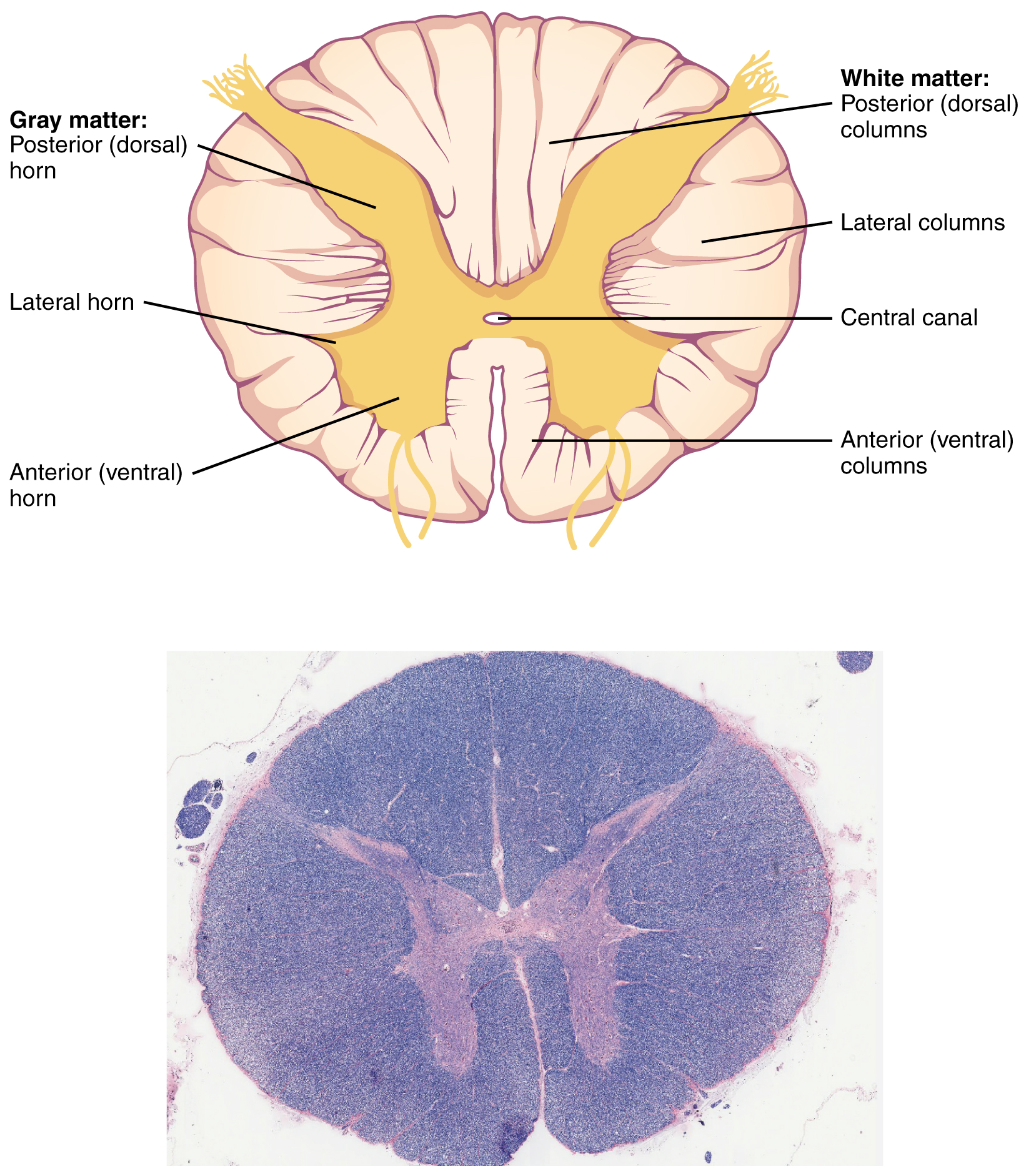
White Columns
Just as the gray matter is separated into horns, the white matter of the spinal cord is separated into columns. Ascending tracts of nervous system fibers in these columns carry sensory information up to the brain, whereas descending tracts carry motor commands from the brain. Looking at the spinal cord longitudinally, the columns extend along its length as continuous bands of white matter. Between the two posterior horns of gray matter are the posterior columns. Between the two anterior horns, and bounded by the axons of motor neurons emerging from that gray matter area, are the anterior columns. The white matter on either side of the spinal cord, between the posterior horn and the axons of the anterior horn neurons, are the lateral columns. The posterior columns are composed of axons of ascending tracts carrying sensory information to the brain. The anterior and lateral columns are composed of many different groups of axons of both ascending and descending tracts—the latter carrying motor commands down from the brain to the spinal cord to control output to the periphery.
External Website

Watch this video to learn about the gray matter of the spinal cord that receives input from fibers of the dorsal (posterior) root and sends information out through the fibers of the ventral (anterior) root. As discussed in this video, these connections represent the interactions of the CNS with peripheral structures for both sensory and motor functions. The cervical and lumbar spinal cords have enlargements as a result of larger populations of neurons. What are these enlargements responsible for?
Glossary
- anterior column
- white matter between the anterior horns of the spinal cord composed of many different groups of axons of both ascending and descending tracts
- anterior horn
- gray matter of the spinal cord containing multipolar motor neurons, sometimes referred to as the ventral horn
- ascending tract
- central nervous system fibers carrying sensory information from the spinal cord or periphery to the brain
- cauda equina
- bundle of spinal nerve roots that descend from the lower spinal cord below the first lumbar vertebra and lie within the vertebral cavity; has the appearance of a horse’s tail
- descending tract
- central nervous system fibers carrying motor commands from the brain to the spinal cord or periphery
- dorsal (posterior) nerve root
- axons entering the posterior horn of the spinal cord
- lateral column
- white matter of the spinal cord between the posterior horn on one side and the axons from the anterior horn on the same side; composed of many different groups of axons, of both ascending and descending tracts, carrying motor commands to and from the brain
- lateral horn
- region of the spinal cord gray matter in the thoracic, upper lumbar, and sacral regions that is the central component of the sympathetic division of the autonomic nervous system
- posterior columns
- white matter of the spinal cord that lies between the posterior horns of the gray matter, sometimes referred to as the dorsal column; composed of axons of ascending tracts that carry sensory information up to the brain
- posterior horn
- gray matter region of the spinal cord in which sensory input arrives, sometimes referred to as the dorsal horn
- posterolateral sulcus
- feature of the posterior spinal cord marking the entry of posterior nerve roots and the separation between the posterior and lateral columns of the white matter
- ventral (anterior) nerve root
- axons emerging from the anterior or lateral horns of the spinal cord
This work, Anatomy & Physiology, is adapted from Anatomy & Physiology by OpenStax, licensed under CC BY. This edition, with revised content and artwork, is licensed under CC BY-SA except where otherwise noted.
Images, from Anatomy & Physiology by OpenStax, are licensed under CC BY except where otherwise noted.
Access the original for free at https://openstax.org/books/anatomy-and-physiology/pages/1-introduction.
