10.7 Smooth Muscle Tissue
Learning Objectives
Understand the structure and function of smooth muscle tissue
By the end of this section, you will be able to:
- Understand the difference between single-unit and multi-unit smooth muscle
- Describe the microanatomy of a smooth muscle cell
- Explain the process of smooth muscle contraction
- Explain how smooth muscle differs from skeletal muscle
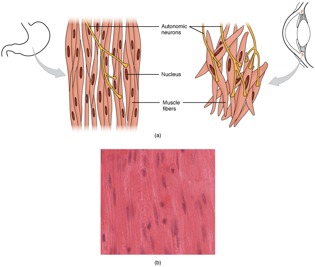
Smooth muscle, so-named because the cells do not have visible striations, is present in the walls of hollow organs (e.g., urinary bladder), lining the blood vessels, and in the eye (e.g., iris) and skin (e.g., erector pili muscle). Smooth muscle displays involuntary control and can be triggered via hormones, neural stimulation by the ANS, and local factors. In certain locations, such as the walls of visceral organs, stretching the muscle can trigger its contraction).
Smooth muscle fibers are spindle-shaped and, unlike skeletal muscle fibers, have a single nucleus; individual cells range in size from 30 to 200 μm. Smooth muscle fibers are often found forming sheets of tissue and function in a coordinated fashion due to the presence of gap junctions between the cells. Termed unitary smooth muscle or visceral muscle, this type of smooth muscle is the most common observed in the human body, forming the walls of hollow organs. Single-unit smooth muscle produces slow, steady contractions that allow substances, such as food in the digestive tract, to move through the body.
Multi-unit smooth muscle, the second type of smooth muscle observed, are composed of cells that rarely possess gap junctions, and thus are not electrically coupled. As a result, contraction does not spread from one cell to the next, but is instead confined to the cell that was originally stimulated. This type of smooth muscle is observed in the large airways to the lungs, in the large arteries, the arrector pili muscles associated with hair follicles, and the internal eye muscles which regulate light entry and lens shape.
External Website

View the University of Michigan WebScope at http://virtualslides.med.umich.edu/Histology/Digestive%20System/Intestines/169_HISTO_40X.svs/view.apml to explore the tissue sample in greater detail.
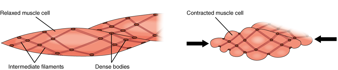
Although smooth muscle cells do not have striations, smooth muscle fibers do have actin and myosin contractile proteins which interact to generate tension. These fibers are not arranged in orderly sarcomeres (hence, no striations) but instead are anchored to dense bodies which are scattered throughout the cytoplasm and anchored to the sarcolemma. A network of intermediate fibers run between the dense bodies providing an internal framework for contractile proteins to work against.
A dense body is analogous to the Z-discs of skeletal muscle, anchoring the thin filaments in position. Calcium ions are supplied primarily from the extracellular environment. T-tubules are absent but small indentations, called calveoli, in the sarcolemma represent locations where there are a high density of calcium channels present to facilitate calcium entry. Sarcoplasmic reticulum is present in the fibers but is less developed than that observed in skeletal muscle.
Because smooth muscle cells do not contain troponin, cross-bridge formation is not regulated by the troponin-tropomyosin complex but instead by the regulatory protein calmodulin. When a smooth muscle cell is stimulated, external Ca++ ions passing through opened calcium channels in the sarcolemma, with additional Ca++ released by the sarcoplasmic reticulum. Calcium binds to calmodulin in the cytoplasm with the Ca++-calmodulin complex then activating an enzyme called myosin (light chain) kinase. Myosin light chain kinase in turn, activates the myosin heads by phosphorylating them (converting ATP to ADP and Pi, with the Pi attaching to the head). The heads can then attach to actin-binding sites and pull on the thin filaments.
When the thin filaments slide past the thick filaments, they pull on the dense bodies, which then pull on the intermediate filaments networks throughout the sarcoplasm. This arrangement causes the entire muscle fiber to contract in a manner whereby the ends are pulled toward the center, causing the midsection to bulge in a corkscrew motion (Figure 10.7.2).
Muscle contraction continues until ATP-dependent calcium pumps actively transport Ca++ ions out of the cell or back into the sarcoplasmic reticulum. However, a low concentration of calcium remains in the sarcoplasm to maintain muscle tone. This remaining calcium keeps the muscle slightly contracted, which is important in certain functions, such as maintaining pressure in blood vessels.
Because most smooth muscles must function for long periods without rest, their power output is relatively low to minimize energy needs. Some smooth muscle can also maintain contractions even as Ca++ is removed and myosin kinase is inactivated/dephosphorylated. This can happen as a subset of cross-bridges between myosin heads and actin, called latch-bridges, keep the thick and thin filaments linked together for a prolonged period, without the need for ATP. This allows for the maintaining of muscle “tone” in smooth muscle that lines arterioles and other visceral organs with very little energy expenditure.
For smooth muscle stimulated by neurons, the axons from autonomic nervous system neurons do not form the highly organized neuromuscular junctions as observed in skeletal muscle. Instead, there is a series of neurotransmitter-filled bulges, called varicosities, along the axon of the neuron feeding the smooth muscle that release neurotransmitters over a wide synaptic cleft. Also, visceral muscle in the walls of the hollow organs (except the heart) contains pacesetter cells. A pacesetter cell can spontaneously trigger action potentials and contractions in the muscle.
Hyperplasia in Smooth Muscle
Similar to skeletal muscle cells, smooth muscle can undergo hypertrophy to increase in size. Unlike other muscle, smooth muscle will also divide quite readily to produce more cells, a process called hyperplasia. This can most evidently be observed in the uterus at puberty, which responds to increased estrogen levels by producing more uterine smooth muscle fibers.
Sections Summary
Smooth muscle is found throughout the body around various organs and tracts. Smooth muscle cells have a single nucleus, and are spindle-shaped. Smooth muscle cells can undergo hyperplasia, mitotically dividing to produce new cells. The smooth cells are nonstriated, but their sarcoplasm is filled with actin and myosin, along with dense bodies in the sarcolemma to anchor the thin filaments and a network of intermediate filaments involved in pulling the sarcolemma toward the fiber’s middle, shortening it in the process. Ca++ ions trigger contraction when they are released from SR and enter through opened voltage-gated calcium channels. Smooth muscle contraction is initiated when the Ca++ binds to intracellular calmodulin, which then activates an enzyme called myosin kinase that phosphorylates myosin heads so they can form the cross-bridges with actin and then pull on the thin filaments. Smooth muscle can be stimulated by pacesetter cells, by the autonomic nervous system, by hormones, spontaneously, or by stretching. The fibers in some smooth muscle have latch-bridges, cross-bridges that cycle slowly without the need for ATP; these muscles can maintain low-level contractions for long periods. Single-unit smooth muscle tissue contains gap junctions to synchronize membrane depolarization and contractions so that the muscle contracts as a single unit. Single-unit smooth muscle in the walls of the viscera, called visceral muscle, has a stress-relaxation response that permits muscle to stretch, contract, and relax as the organ expands. Multiunit smooth muscle cells do not possess gap junctions, and contraction does not spread from one cell to the next.
Review Questions
Critical Thinking Questions
1. Why can smooth muscles contract over a wider range of resting lengths than skeletal and cardiac muscle?
2. Describe the differences between single-unit smooth muscle and multiunit smooth muscle.
Glossary
- calmodulin
- regulatory protein that facilitates contraction in smooth muscles
- dense body
- sarcoplasmic structure that attaches to the sarcolemma and shortens the muscle as thin filaments slide past thick filaments
- hyperplasia
- process in which one cell splits to produce new cells
- latch-bridges
- subset of a cross-bridge in which actin and myosin remain locked together
- pacesetter cell
- cell that triggers action potentials in smooth muscle
- stress-relaxation response
- relaxation of smooth muscle tissue after being stretched
- varicosity
- enlargement of neurons that release neurotransmitters into synaptic clefts
- visceral muscle
- smooth muscle found in the walls of visceral organs
Solutions
Answers for Critical Thinking Questions
- Smooth muscles can contract over a wider range of resting lengths because the actin and myosin filaments in smooth muscle are not as rigidly organized as those in skeletal and cardiac muscle.
- Single-unit smooth muscle is found in the walls of hollow organs; multiunit smooth muscle is found in airways to the lungs and large arteries. Single-unit smooth muscle cells contract synchronously, they are coupled by gap junctions, and they exhibit spontaneous action potential. Multiunit smooth cells lack gap junctions, and their contractions are not synchronous.
This work, Anatomy & Physiology, is adapted from Anatomy & Physiology by OpenStax, licensed under CC BY. This edition, with revised content and artwork, is licensed under CC BY-SA except where otherwise noted.
Images, from Anatomy & Physiology by OpenStax, are licensed under CC BY except where otherwise noted.
Access the original for free at https://openstax.org/books/anatomy-and-physiology/pages/1-introduction.
