12.2 Nervous Tissue
Learning Objectives
By the end of this section, you will be able to:
Explain how neurons and glial cells work together to perform and support the nervous system functions.
- Describe the basic structure of a neuron and how these structures function in a neuron
- Identify the different types of neurons on the basis of shape
- List the glial cells of the CNS and describe their function
- List the glial cells of the PNS and describe their function
Nervous tissue is composed of two types of cells, neurons and glial cells. Neurons are responsible for the computation and communication that the nervous system provides. They are electrically active and release chemical signals to communicate between each other and with target cells. Glial cells, or glia or neuroglia, are much smaller than neurons and play a supporting role for nervous tissue. Glial cells maintain the extracellular environment around neurons, improve signal conduction in neurons and protect them from pathogens. Ongoing research also suggests that glial cell number matches neuron number and that they even can send signals themselves.
Neuron Anatomy
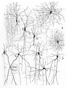
Neurons are nucleated cells with specialized structural properties. Some neurons have a single long extension (axon) that reaches great distances, others are very small, star shaped cells without obvious axons (See Figure 12.2.1 – add to image the term axon, reference cells without one).
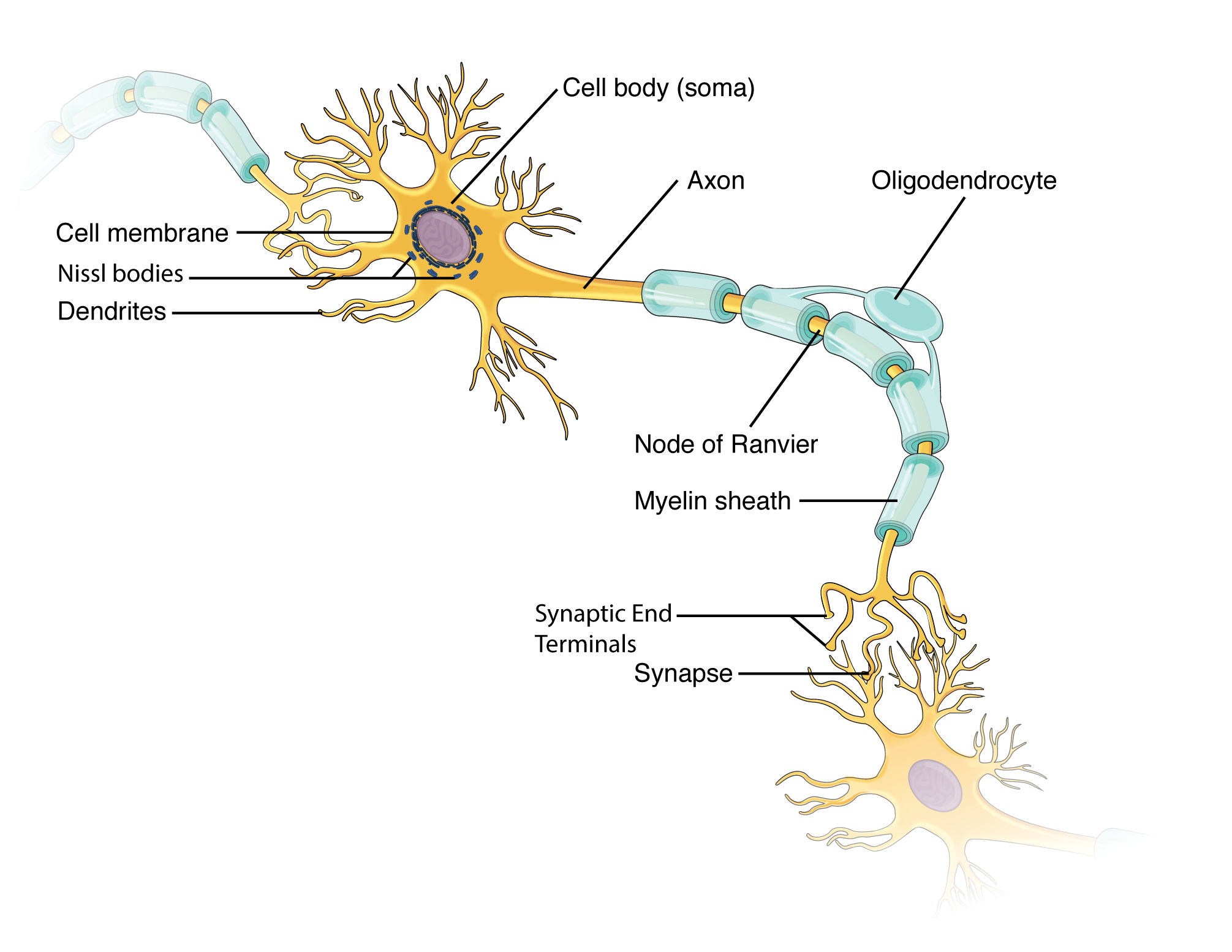
Though neuron shapes vary greatly, every neuron houses its nucleus in a region known as the cell body (also called soma) from which cellular activity like repair or cell membrane recycling is controlled. Associated with the nucleus, neurons also have many rough endoplasmic reticula, called Nissl bodies (these can be seen in neurons using a light microscope). The nucleus, Nissl bodies and golgi apparatuses together produce the many ion channels and pumps that reside in the cell membrane. These transmembrane proteins are neccessary for neurons to send electrical signals (graded potentials and action potentials, see section 12.4). In addition, neurons consume much ATP and typically have many mitochondria.
In figure 12.2.2, the cell body shows both many short projections and one long projection emerging from the cell body. These short projections are dendrites which receive most of the input from other neurons or stimuli in the extracellular environment; the location of the dendrites on the neuron marks the receptive region of the neuron. Dendrites are usually highly branched processes, providing locations for other neurons to communicate with the neuron. Neurons have polarity—meaning that information flows in one direction through the neuron. In the figure 12.2.2 neuron, information flows from the dendrites, across the cell body, and down the large axon emerging from the cell body at the axon hillock (axon hillock is an anatomical term to describe where the cell body and axon meet). The first section of the axon where an action potential is generated is called the initial segment. In multipolar and bipolar neurons, the initial segment is found at the axon hillock (see Figure 12.2.3). However, in unipolar neurons, the initial segment is not found at the axon hillock, and can actually be located many inches or even a few feet from it near the dendrites (see Figure 12.2.3)! However, in unipolar neurons, the initial segment is not found at the axon hillock, and can actually be located many inches or even a few feet from it! Often axons are wrapped by myelin sheaths, leaving exposed sections (node of Ranvier) between segments of myelin. Myelin is produced by oligodendrocytes (glial cells) in the CNS and Schwann cells in the PNS; it acts as electrical insulation, speeding information conduction down the neuron. Once information reaches the terminal end of this neuron, it is transferred to another cell. The site of communication between a neuron and its target cell is called a synapse. The terminal end has several branches, each with a synaptic end bulb to store chemicals needed for communication with the next cell. Figure 12.2.2 shows the relationship of these parts to one another.
External Website

Visit this site (link not working as of 10/20/2021) to learn about how nervous tissue is composed of neurons and glial cells. Neurons are dynamic cells with the ability to make a vast number of connections, to respond incredibly quickly to stimuli, and to initiate movements on the basis of those stimuli. They are the focus of intense research because failures in physiology can lead to devastating illnesses. Why are neurons only found in animals? Based on what this article says about neuron function, why wouldn’t they be helpful for plants or microorganisms?
Types of Neurons
There are trillions of neurons in the nervous system and cell shape can vary widely. Three common shapes of neurons are shown in Figure 12.2.3.
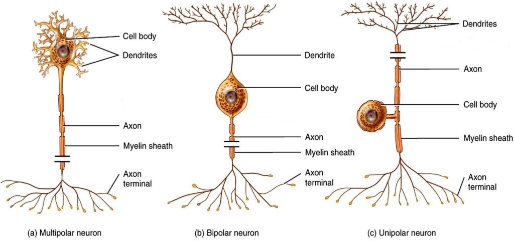
Multipolar neurons have multiple processes emerging from their cell bodies (hence their name, multipolar). They have dendrites attached to their cell bodies and often, one long axon. Motor neurons are multipolar neurons, as are many neurons of the CNS.
Bipolar cells have two processes, which extend from each end of the cell body, opposite to each other. One is the axon and one the dendrite. Bipolar cells are not very common. They are found mainly in the olfactory epithelium (where smell stimuli are sensed), and as part of the retina in the eye.
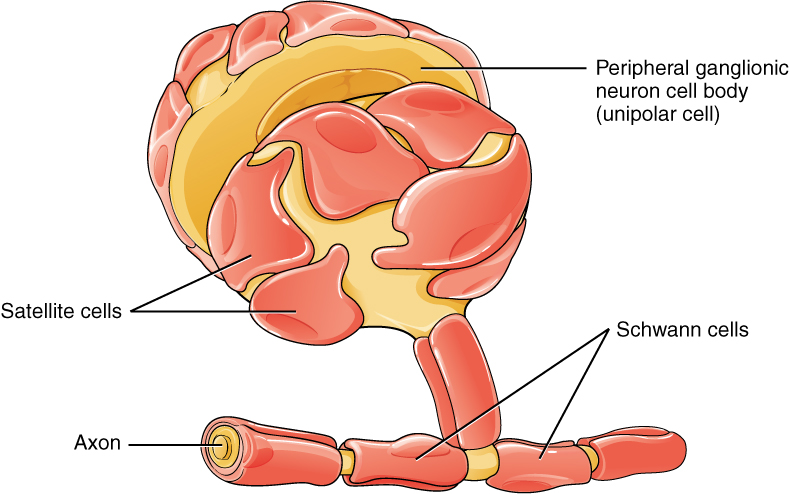
Unipolar cells have one long axon emerging from the cell body, with the cell body located between the two ends, and off to the side. At one end of the axon are dendrites, and at the other end, the axon forms synaptic connections with a target cell. Unipolar cells are exclusively sensory neurons and have their dendrites in the periphery where they detect stimuli. Their cell bodies are typically found in ganglia of the peripheral nervous system.
Glial Cells
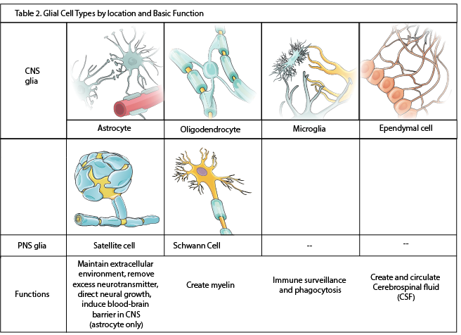
There are six types of glial cells. Four of them are found in the CNS and two are found in the PNS. Table 12.1 outlines some common characteristics and functions.
Glial Cells of the CNS
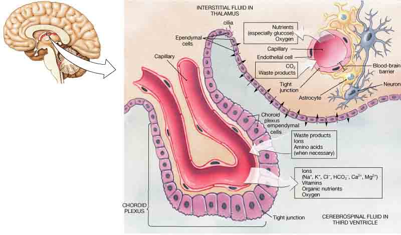
One cell providing support to neurons of the CNS is the astrocyte, so named because it appears to be star-shaped under the microscope (astro- = “star”, cyte = “cell”). Astrocytes have many processes extending from their main cell body (not axons or dendrites like neurons, just cell extensions). Those processes extend to interact with neurons, blood vessels, or the connective tissue covering the CNS (Figure 12.2.4). Generally, they are supporting cells for the neurons in the central nervous system. Some ways in which they support neurons in the central nervous system are by maintaining the concentration of chemicals in the extracellular space, removing excess signaling molecules, reacting to tissue damage, and inducing to the blood-brain barrier (BBB). The blood-brain barrier is a protective physiological barrier that keeps many substances that circulate in the blood from getting into the central nervous system, restricting what can cross from circulating blood into the CNS. Usually, blood vessels are leaky because there are gaps between the cells of the vessel walls. These gaps permit rapid movement of molecules out of the blood into the extracellular space around tissue cells, delivering nutrients and hormones. However, the neurons of the brain may be affected by rapid, regular changes in extracellular concentrations preventing signal transmission. To prevent such fluctuations, astrocytes release compounds to the blood vessels, inducing tight junctions between the otherwise leaky blood vessel cells. When the BBB is intact, nutrient molecules, such as glucose or amino acids, must now pass through the vessel cells of the BBB by transcellular processes (using membrane proteins). Small, fat soluble molecules (respiratory gases, alcohol) are able simply diffuse through the cell membranes, but other large, water soluble molecules cannot. The highly restrictive permeability of the BBB may restrict drug delivery to the CNS. Pharmaceutical companies are challenged to design drugs that can cross the BBB as well as have an effect on the nervous system.
Also found in CNS tissue is the oligodendrocyte, sometimes called just “oligo,” which is the glial cell type that insulates axons in the CNS. The name means “cell of a few branches” (oligo- = “few”; dendro- = “branches”; -cyte = “cell”). There are a few processes that extend from the cell body. Each one reaches out and surrounds an axon to insulate it in myelin. One oligodendrocyte will provide the myelin for multiple axon segments, either for the same axon or for separate axons. The function of myelin will be discussed below.
Microglia are, as the name implies, smaller than most of the other glial cells. Ongoing research into these cells, although not entirely conclusive, suggests that they may originate as white blood cells, called macrophages, that become part of the CNS during early development. While their origin is not conclusively determined, their function is related to what macrophages do in the rest of the body. When macrophages encounter diseased or damaged cells in the rest of the body, they ingest and digest those cells or the pathogens that cause disease. Microglia are the cells in the CNS that can do this in normal, healthy tissue, and they are therefore also referred to as CNS-resident macrophages.
Ependymal cells filter blood to make cerebrospinal fluid (CSF), the fluid that circulates through the CNS. CSF is needed in the brain to provide nutrients, remove wastes and create a stable extracellular environment because the BBB is so restrictive. In each of the brain cavities (ventricles), ependymal cells surround the blood vessels forming choroid plexuses. These choroid plexuses filter specific components of the blood to produce cerebrospinal fluid. Everyday they produce enough CSF to fill a pint glass! Though the BBB is absent in the choroid plexuses, the ependymal cells there are connected to each other by tight connections, forming a highly restrictive boundary. More ependymal cells line the ventricles and use their cilia to help move the CSF through the ventricular space. The relationship of these glial cells to the structure of the CNS is seen in Figure 12.2.4.
Glial Cells of the PNS
One of the two types of glial cells found in the PNS is the satellite cell. Satellite cells surround the cell bodies of neurons in the PNS. They provide support, performing similar functions in the periphery as astrocytes do in the CNS—except, of course, for establishing the BBB.
The second type of glial cell is the Schwann cell, which insulate axons with myelin in the periphery. Schwann cells are different than oligodendrocytes in that a Schwann cell wraps around a portion of only one axon segment and no others. Oligodendrocytes have processes that reach out to multiple axon segments, whereas the entire Schwann cell surrounds just one axon segment. The nucleus and cytoplasm of the Schwann cell are on the edge of the myelin sheath. The relationship of these two types of glial cells to ganglia and nerves in the PNS is seen in Figure 12.2.5.
Myelin
Oligodendrocytes in the CNS and Schwann cells in the PNS provide myelin. Whereas the manner in which either cell is associated with the axon segment, or segments, that it insulates is different, the means of myelinating an axon segment is mostly the same in the two situations. Myelin is a lipid-rich sheath that surrounds the axon and by doing so creates a myelin sheath that facilitates the transmission of electrical signals along the axon. Simply, myelinated axons send signals faster than unmyelinated axons. The lipids of myelin are essentially the phospholipids of the glial cell membrane. Myelin, however, is more than just the membrane of the glial cell. It also includes important proteins that are integral to that membrane. Some of the proteins help to hold the layers of the glial cell membrane closely together.
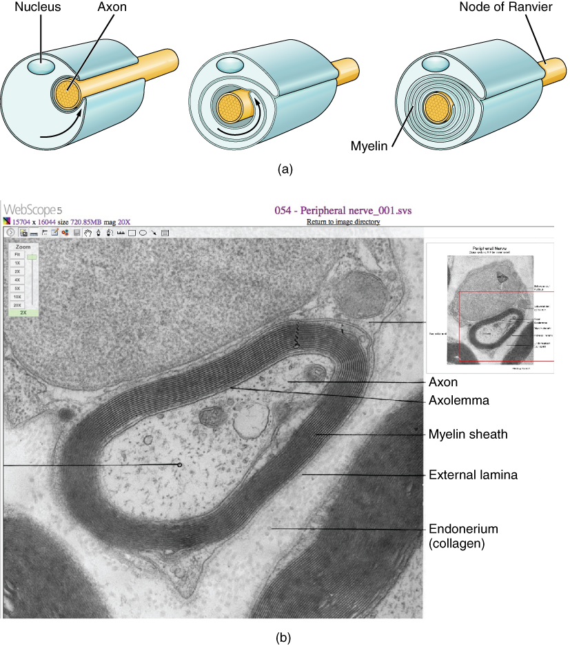
The appearance of the myelin sheath can be thought of as similar to the pastry wrapped around a hot dog for “pigs in a blanket” or a similar food. The glial cell is wrapped around the axon several times with little to no cytoplasm between the glial cell layers. For oligodendrocytes, the rest of the cell is separate from the myelin sheath as a cell process extends back toward the cell body. A few other processes provide the same insulation for other axon segments in the area. For Schwann cells, the outermost layer of the cell membrane contains cytoplasm and the nucleus of the cell as a bulge on one side of the myelin sheath. During development, the glial cell is loosely or incompletely wrapped around the axon (Figure 12.2.6a). The edges of this loose enclosure extend toward each other, and one end tucks under the other. The inner edge wraps around the axon, creating several layers, and the other edge closes around the outside so that the axon is completely enclosed.
External Website

View the University of Michigan WebScope at https://histologyslides.med.umich.edu/Histology/EMsmallCharts/3%20Image%20Scope%20finals/054%20-%20Peripheral%20nerve_001.htm to see an electron micrograph of a cross-section of a myelinated nerve fiber.
Myelin sheaths can extend for one or two millimeters, depending on the diameter of the axon. Axon diameters can be as small as 1 to 20 micrometers. Because a micrometer is 1/1000 of a millimeter, this means that the length of a myelin sheath can be 100–1000 times the diameter of the axon. Figure 12.2.2, Figure 12.2.4, and Figure 12.2.5 show the myelin sheath surrounding an axon segment, but are not to scale. If the myelin sheath were drawn to scale, the neuron would have to be immense—possibly covering an entire wall of the room in which you are sitting.
Disorders of the…Nervous Tissue
Several diseases can result from the demyelination of axons. The causes of these diseases are not the same; some have genetic causes, some are caused by pathogens, and others are the result of autoimmune disorders. Though the causes are varied, the results are largely similar. The myelin insulation of axons is compromised, making electrical signaling slower. In some cases, signaling stops, preventing muscles from responding and causing paralysis.
Multiple sclerosis (MS) is one such disease. It is an example of an autoimmune disease. The antibodies produced by lymphocytes (a type of white blood cell) mark myelin as something that should not be in the body. This causes inflammation and the destruction of the myelin in the central nervous system. As the insulation around the axons is destroyed by the disease, scarring occurs. This is where the name of the disease comes from; sclerosis means hardening of tissue, as occurs in a scar. Multiple scars are found in the white matter of the brain and spinal cord. Control of the skeletal and smooth musculature is compromised, affecting not only movement, but also control of organs such as the bladder.
Guillain-Barré (pronounced gee-YAN bah-RAY) syndrome is an example of a demyelinating disease of the peripheral nervous system. It is also the result of an autoimmune reaction, but the inflammation is in peripheral nerves. Sensory symptoms or motor deficits are common, and autonomic failures can lead to changes in the heart rhythm or a drop in blood pressure, especially when standing, which causes dizziness.
Chapter Review
Nervous tissue contains two major cell types, neurons and glial cells. Neurons are responsible for communication through electrical signals. Glial cells are supporting cells, allowing neuron function.
Though neuron shape varies, neurons are polarized cells, based on the flow of electrical signals along their membrane. In multipolar neurons, dendrites receive signals and pass them to the cell body; signals then propagate along the axon towards the terminal end that synapses with a target cell. Myelin on axons speeds signal conduction and is provided by different glial cells in the CNS and PNS.
The nervous system has several types of glial cells, categorized by the anatomical division in which they are found. In the CNS, astrocytes, oligodendrocytes, microglia, and ependymal cells perform different functions that support neurons. Astrocytes maintain the chemical environment around neurons and are crucial for regulating the blood-brain barrier. Oligodendrocytes myelinate neurons, microglia act as phagocytes and play a role in immune surveillance. Ependymal cells filter blood to produce cerebrospinal fluid (CSF). CSF circulates through the CNS proving nutrients and removing waste. In the PNS, satellite cells maintain the extracellular environment around cell bodies and Schwann cells insulate peripheral axons.
Interactive Link Questions
Visit this site (link does not work as 11/03/2021) to learn about how nervous tissue is composed of neurons and glial cells. The neurons are dynamic cells with the ability to make a vast number of connections and to respond incredibly quickly to stimuli and to initiate movements based on those stimuli. They are the focus of intense research as failures in physiology can lead to devastating illnesses. Why are neurons only found in animals? Based on what this article says about neuron function, why wouldn’t they be helpful for plants or microorganisms?
Neurons enable thought, perception, and movement. Plants do not move, so they do not need this type of tissue. Microorganisms are too small to have a nervous system. Many are single-celled, and therefore have organelles for perception and movement.
View the University of Michigan WebScope at https://histologyslides.med.umich.edu/Histology/EMsmallCharts/3%20Image%20Scope%20finals/054%20-%20Peripheral%20nerve_001.htm to see an electron micrograph of a cross-section of a myelinated nerve fiber. The axon contains microtubules and neurofilaments, bounded by a plasma membrane known as the axolemma. Outside the plasma membrane of the axon is the myelin sheath, which is composed of the tightly wrapped plasma membrane of a Schwann cell. What aspects of the cells in this image react with the stain that makes them the deep, dark, black color, such as the multiple layers that are the myelin sheath?
Exercises
Critical Thinking Questions
1. Multiple sclerosis is a demyelinating disease affecting the central nervous system. What type of cell would be the most likely target of this disease? Why?
2. Suppose a unipolar neuron has half of its axon in the CNS and the other half in the PNS. If this neuron is fully myelinated, what cells would be involved and where?
Glossary
- astrocyte
- glial cell type of the CNS that provides support for neurons and maintains the blood-brain barrier
- axon
- single process of the neuron that carries an electrical signal (action potential) away from the cell body toward a target cell
- axon hillock
- region of the neuron cell body that gives rise to the axon
- axon terminal (terminal end)
- end of the axon, where there are usually several branches extending toward the target cell
- bipolar neuron
- shape of a neuron with two processes extending from the neuron cell body—the axon and one dendrite
- blood-brain barrier (BBB)
- physiological barrier between the circulatory system and the central nervous system that establishes a privileged blood supply, restricting the flow of substances into the CNS
- cerebrospinal fluid (CSF)
- circulatory medium within the CNS that is produced by ependymal cells in the choroid plexus filtering the blood
- choroid plexus
- specialized structure containing ependymal cells that line blood capillaries and filter blood to produce CSF in the four ventricles of the brain
- dendrite
- one of many branchlike processes that extends from the neuron cell body and functions as a contact for incoming signals (synapses) from other neurons or sensory cells
- ependymal cell
- glial cell type in the CNS responsible for producing cerebrospinal fluid
- glial cell
- one of the various types of neural tissue cells responsible for maintenance of the tissue, and largely responsible for supporting neurons
- initial segment
- first part of axon where the electrical signals known as action potentials are generated.
- microglia
- glial cell type in the CNS that serves as the resident component of the immune system
- multipolar
- shape of a neuron that has multiple processes—the axon and two or more dendrites
- myelin
- lipid-rich insulating substance surrounding the axons of many neurons, allowing for faster transmission of electrical signals
- myelin sheath
- lipid-rich layer of insulation that surrounds an axon, formed by oligodendrocytes in the CNS and Schwann cells in the PNS; facilitates the transmission of electrical signals
- neuron
- neural tissue cell that is primarily responsible for generating and propagating electrical signals into, within, and out of the nervous system
- node of Ranvier
- gap between two myelinated regions of an axon, allowing for strengthening of the electrical signal as it propagates down the axon
- oligodendrocyte
- glial cell type in the CNS that provides the myelin insulation for axons in tracts
- satellite cell
- glial cell type in the PNS that provides support for neurons in the ganglia
- Schwann cell
- glial cell type in the PNS that provides the myelin insulation for axons in nerves
- soma (cell body)
- in neurons, that portion of the cell that contains the nucleus
- synapse
- site of communication between a neuron and another cell
- synaptic end bulb
- swelling at the end of an axon where neurotransmitter molecules are released onto a target cell across a synapse
- unipolar
- shape of a neuron which has only one process that includes both the axon and dendrite
- ventricle
- central cavity within the brain where CSF is produced and circulates
Solutions
Answers for Critical Thinking Questions
- The disease would target oligodendrocytes. In the CNS, oligodendrocytes provide the myelin for axons.
- Unipolar neurons have a long axon. If half is in the CNS, that half will be myelinated by oligodendrocytes. The half in the PNS will be myelinated by Schwann cells.
This work, Anatomy & Physiology, is adapted from Anatomy & Physiology by OpenStax, licensed under CC BY. This edition, with revised content and artwork, is licensed under CC BY-SA except where otherwise noted.
Images, from Anatomy & Physiology by OpenStax, are licensed under CC BY except where otherwise noted.
Access the original for free at https://openstax.org/books/anatomy-and-physiology/pages/1-introduction.
