11.1 Describe the roles of agonists, antagonists and synergists
Learning Objectives
By the end of this section, you will be able to identify the following:
Compare and contrast agonist and antagonist muscles
The moveable end of the muscle that attaches to the bone being pulled is called the muscle’s insertion, and the end of the muscle attached to a fixed (stabilized) bone is called the origin. Muscle pull rather than push. Upon activation, the muscle pulls the insertion toward the origin.
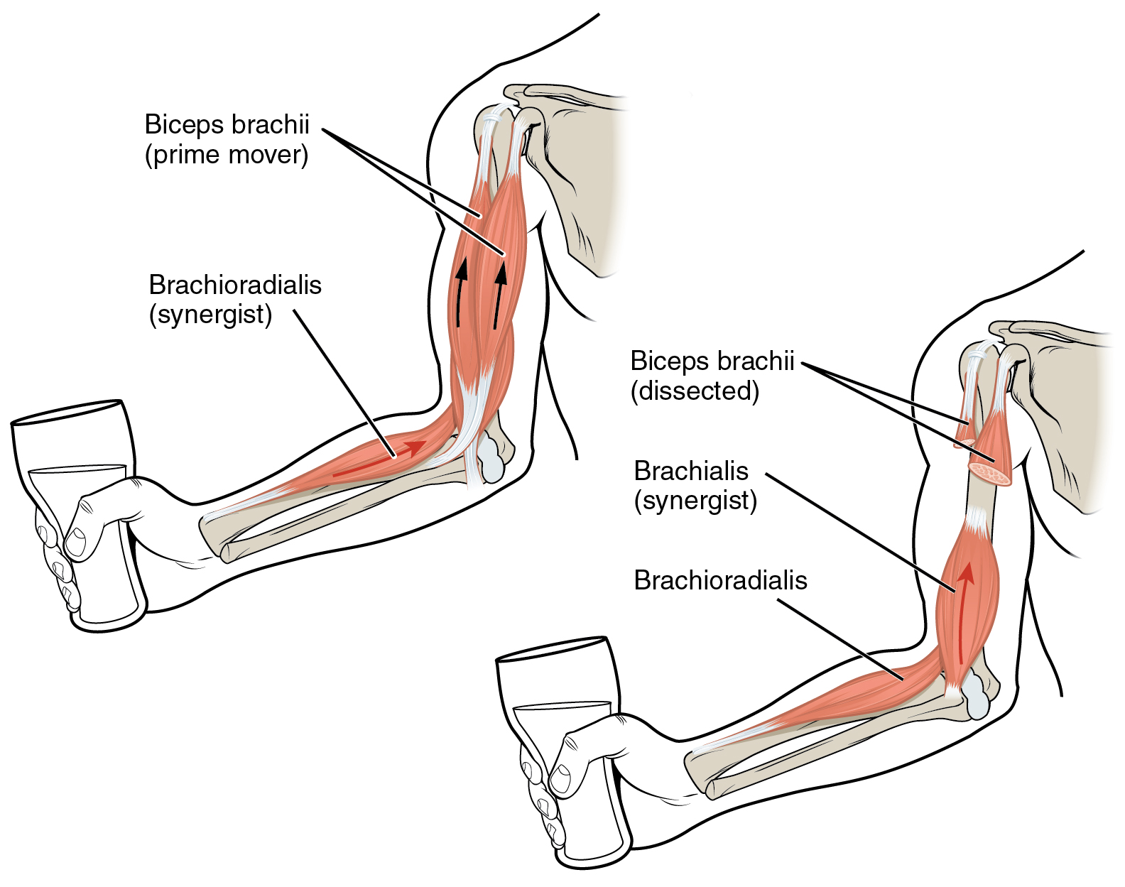
Although a number of muscles may be involved in an action, the principal muscle involved is called the prime mover, or agonist. During forearm flexion, for example lifting a cup, a muscle called the biceps brachii is the prime mover. Because it can be assisted by the brachialis, the brachialis is called a synergist in this action (Figure 11.1.1). A synergist can also be a fixator that stabilizes the muscle’s origin.
A muscle with the opposite action of the prime mover is called an antagonist. Antagonists play two important roles in muscle function: (1) they maintain body or limb position, such as holding the arm out or standing erect; and (2) they control rapid movement, as in shadow boxing without landing a punch or the ability to check the motion of a limb.
For example, to extend the leg at the knee, a group of four muscles called the quadriceps femoris in the anterior compartment of the thigh are activated (and would be called the agonists of leg extension at the knee). A set of antagonists called the hamstrings in the posterior compartment of the thigh are activated to slow or stop the movement.
These terms are reversed for the opposite action, flexion of the leg at the knee. In this case the hamstrings would be called the agonists and the quadriceps femoris would be called the antagonists.
There are also muscles that do not pull against the skeleton for movements such as the muscles of facial expressions. The insertions and origins of facial muscles are in the skin, so that certain individual muscles contract to form a smile or frown, form sounds or words, and raise the eyebrows. There also are skeletal muscles in the tongue, and the external urinary and anal sphincters that allow for voluntary regulation of urination and defecation, respectively. There are four helpful rules that can be applied to all major joints except the ankle and knee because the lower extremity is rotated during development. For example, in the case of the knee, muscles of the posterior thigh cause knee flexion and anterior thigh muscles cause knee extension, which is opposite of the rules stated below for most other joints.
- A muscle that crosses the anterior side of a joint results in flexion, which results in a decrease in joint angle with movement. For example, the anterior arm muscles cause elbow flexion. FIGURE OF ISOLATED BICEPS BRACHII. Like Figure 10.15c in Marieb-11e.
- A muscle that crosses the posterior side of a joint results in extension, which results in an increase in joint angle with movement. For example, the muscles in the posterior arm cause elbow extension. FIGURE OF ISOLATED TRICEPS BRACHII. Like Figure 10.15b in Marieb-11e.
- A muscle that crosses the lateral side of a joint results in abduction, which results in the body part moving away from the midline of the body. For example, the deltoid muscle on the lateral side of the upper arm causes abduction of the shoulder. INSERT FIGURE LIKE FOCUS FIGURE 10.1c IN MARIEB-11E
- A muscle that crosses the medial side of a joint results in adduction, which results in the upper or lower extremity moving toward the midline of the body. For example, the teres major muscle, on the medial side of the arm causes shoulder abduction. INSERT FIGURE LIKE FOCUS FIGURE 10.1d IN MARIEB-11E.
Chapter Review
Skeletal muscles each have an origin and an insertion. The end of the muscle that attaches to the bone being pulled is called the muscle’s insertion and the end of the muscle attached to a fixed, or stabilized, bone is called the origin. The muscle primarily responsible for a movement is called the prime mover, and muscles that assist in this action are called synergists. A synergist that makes the insertion site more stable is called a fixator. Meanwhile, a muscle with the opposite action of the prime mover is called an antagonist.
Review Questions
Glossary
- abduction
- the body part moves away from the midline of the body
- adduction
- the body part moves toward the midline of the body
- agonist
- (also, prime mover) muscle whose contraction is responsible for producing a particular motion
- antagonist
- muscle that opposes the action of an agonist
- extension
- an increase in joint angle with movement
- fixator
- synergist that assists an agonist by preventing or reducing movement at another joint, thereby stabilizing the origin of the agonist
- flexion
- a decrease in joint angle with movement
- insertion
- end of a skeletal muscle that is attached to the structure (usually a bone) that is moved when the muscle contracts
- origin
- end of a skeletal muscle that is attached to another structure (usually a bone) in a fixed position
- prime mover
- (also, agonist) principle muscle involved in an action
- synergist
- muscle whose contraction helps a prime mover in an action
This work, Anatomy & Physiology, is adapted from Anatomy & Physiology by OpenStax, licensed under CC BY. This edition, with revised content and artwork, is licensed under CC BY-SA except where otherwise noted.
Images, from Anatomy & Physiology by OpenStax, are licensed under CC BY except where otherwise noted.
Access the original for free at https://openstax.org/books/anatomy-and-physiology/pages/1-introduction.
