4.0 Introduction
Chapter Objectives
After studying this chapter, you will be able to:
4.1 – Identify the main tissue types and discuss their roles in the human body.
4.2 – Describe the structural characteristics of the various epithelial tissues and how these characteristics enable their functions.
4.3 – Describe the structural characteristics of the various connective tissues and how these characteristics enable their functions.
4.4 – Describe the characteristics of muscle tissue and how these dictate muscle function.
4.5 – Describe the characteristics of nervous tissue and how these enable the unique functions of nervous tissue.
4.6 – Describe the process of tissue response to injury.
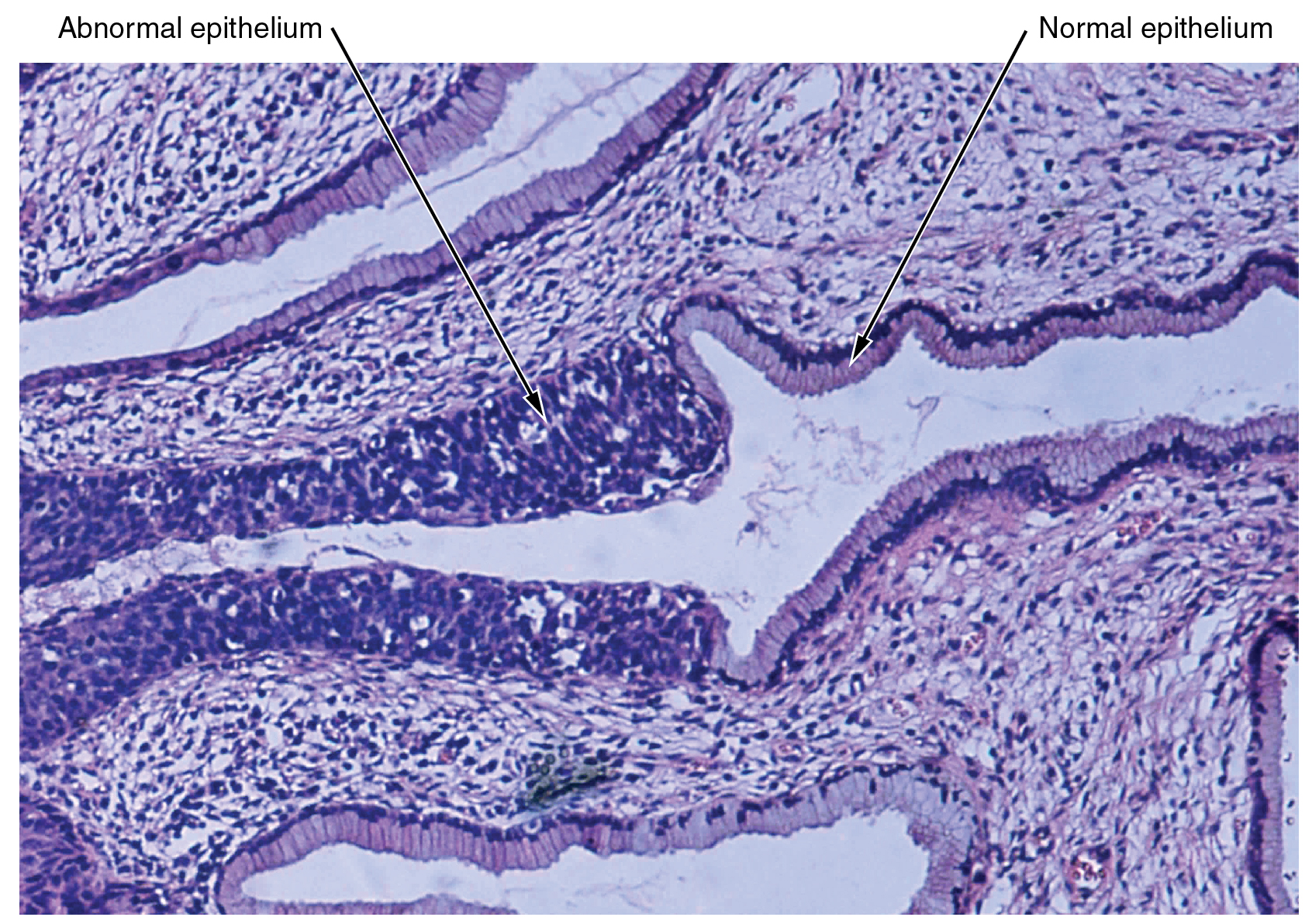
The cells found in the human body contain essentially the same internal structures yet they vary enormously in shape and function. The variation in cells is not randomly distributed throughout the body, rather, they occur in organized layers. Such aggregations of cells that are similar in structure and work together to perform a specialized function are referred to as tissues. The micrograph that opens this chapter shows the high degree of organization among different types of cells in the tissue of the cervix. You can also see how that organization breaks down when cancer takes over the regular mitotic functioning of a cell.
The human body starts as a single cell at fertilization. As this fertilized egg divides, it gives rise to trillions of cells, each built from the same blueprint, but organizing into tissues and becoming irreversibly committed to a developmental pathway.
This work, Anatomy & Physiology, is adapted from Anatomy & Physiology by OpenStax, licensed under CC BY. This edition, with revised content and artwork, is licensed under CC BY-SA except where otherwise noted.
Images, from Anatomy & Physiology by OpenStax, are licensed under CC BY except where otherwise noted.
Access the original for free at https://openstax.org/books/anatomy-and-physiology/pages/1-introduction.
