25.1 Internal and External Anatomy of the Kidney
Learning Objectives
By the end of this section, you will be able to:
Describe the macroscopic and microscopic anatomy of the kidney.
- Describe the external structure of the kidney, including its location, support structures, and covering
- Identify the major internal divisions and structures of the kidney
- Identify the major blood vessels associated with the kidney and trace the path of blood through the kidney
- Identify the major structures and subdivisions of the nephron and describe them histologically
External Website

There have never been sufficient kidney donations to provide a kidney to each person needing one. Watch this video to learn about the TED (Technology, Entertainment, Design) Conference held in March 2011. In this video, Dr. Anthony Atala discusses a cutting-edge technique in which a new kidney is “printed.” The successful utilization of this technology is still several years in the future, but imagine a time when you can print a replacement organ or tissue on demand.
External Anatomy
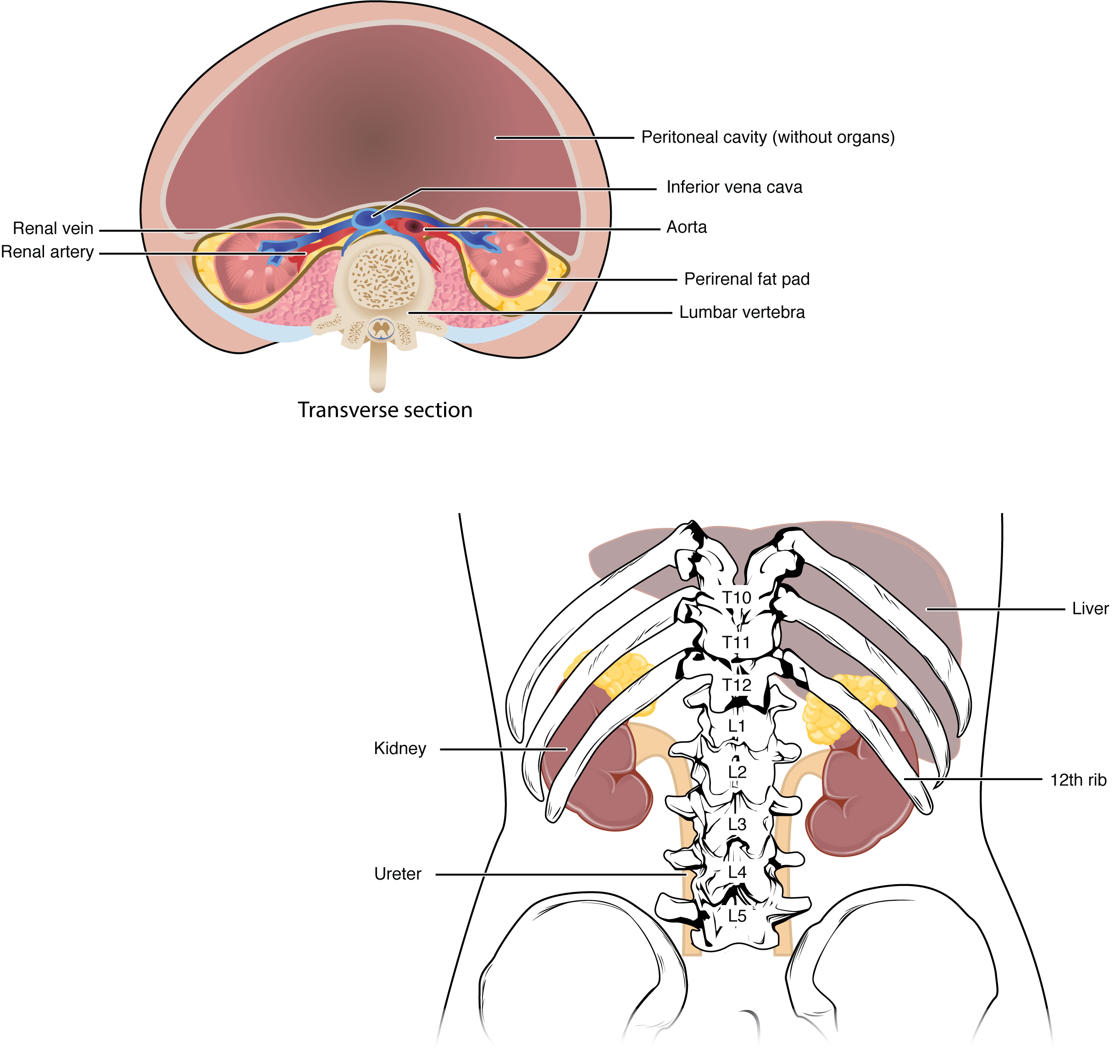
The paired kidneys lie on either side of the spine in the retroperitoneal space between the parietal peritoneum and the posterior abdominal wall, well protected by muscle, fat, and ribs. The left kidney is located at about the T12 to L3 vertebrae, whereas the right is lower due to slight displacement by the liver. Upper portions of the kidneys are somewhat protected by the eleventh and twelfth ribs (Figure 25.1.1). Each kidney weighs about 125–175 g in males and 115–155 g in females. They are about 11–14 cm in length, 6 cm wide, and 4 cm thick, and are directly covered by a fibrous capsule composed of dense, irregular connective tissue that helps to hold their shape and protect them. This capsule is covered by a shock-absorbing layer of adipose tissue called the renal fat pad, which in turn is encompassed by a tough renal fascia. The fascia and, to a lesser extent, the overlying peritoneum serve to firmly anchor the kidneys to the posterior abdominal wall in a retroperitoneal position.
Internal Anatomy
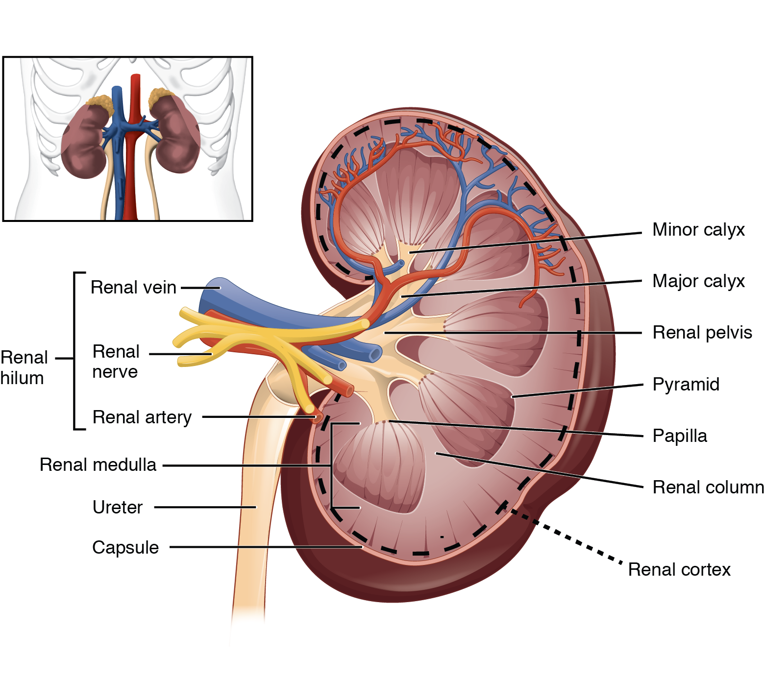
A frontal section through the kidney reveals an outer region called the renal cortex and an inner region called the renal medulla (Figure 25.1.2). In the medulla, 5-8 renal pyramids are separated by connective tissue renal columns. Each pyramid creates urine and terminates into a renal papilla. Each renal papilla drains into a collecting pool called a minor calyx; several minor calyces connect to form a major calyx; all major calyces connect to the single renal pelvis which connects to the ureter.
Blood Supply of the Kidney & Nephrons
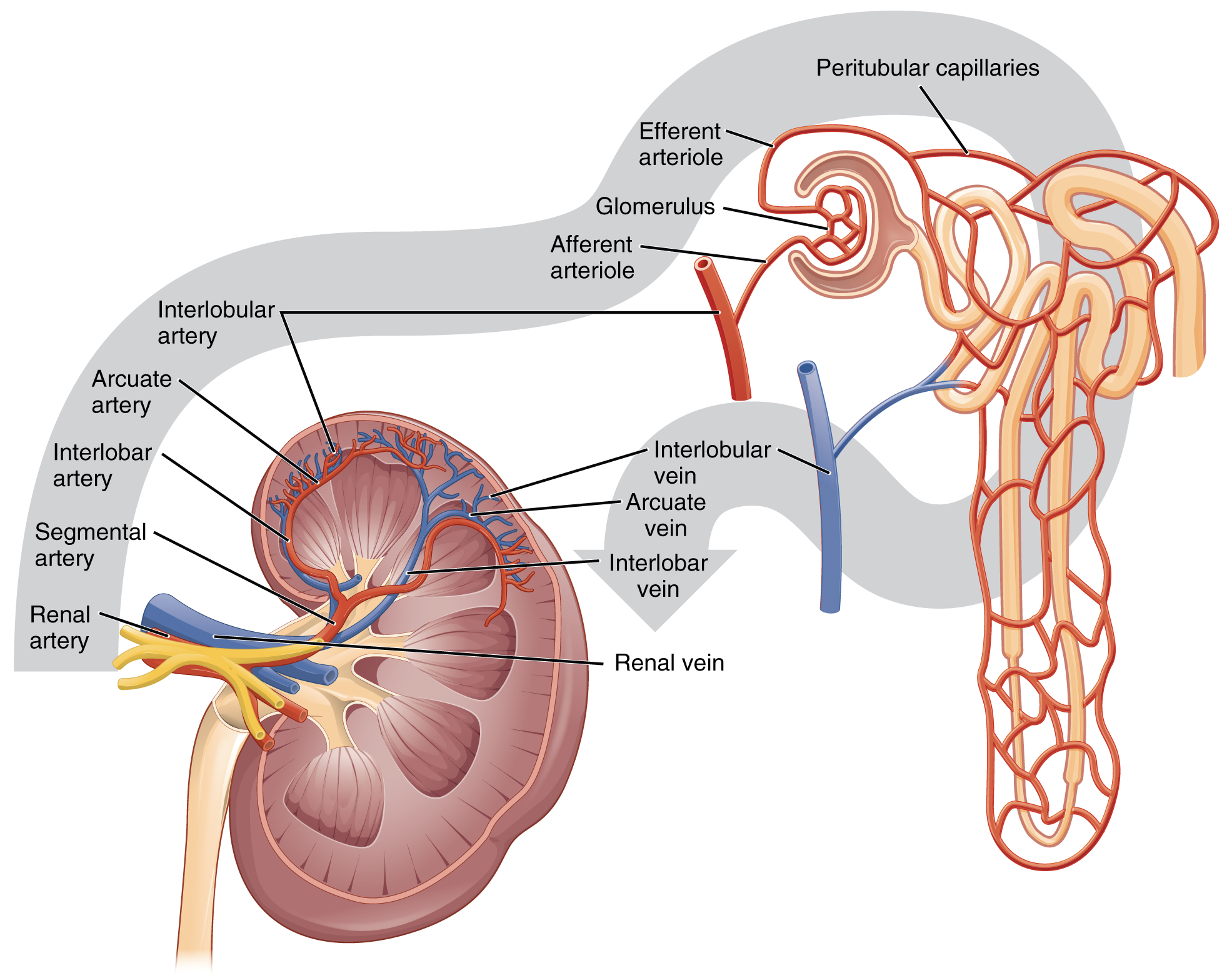
The kidneys are well vascularized and receive about 25 percent of the cardiac output at rest. Blood enters the kidney via the paired renal arteries that form directly from the descending aorta and each enters the kidney at the renal hila. Once in the kidney, each renal artery first divides into segmental arteries, followed by further branching to form interlobar arteries that pass through the renal columns to reach the cortex (Figure 25.1.3). The interlobar arteries, in turn, branch into arcuate arteries, cortical radiate arteries, and then into afferent arterioles. The afferent arterioles deliver blood into a modified capillary bed called the glomerulus which is a component of the “functional unit” of the kidney called the nephron. There are about 1.3 million nephrons in each kidney and they function to filter the blood. Once the nephrons have filtered the blood, renal veins return blood directly to the inferior vena cava. A portal system is formed when the blood flows from the glomerulus to the efferent arteriole through a second capillary bed, the peritubular capillaries (and vasa recta), surrounding the proximal and distal convoluted tubules and the loop of Henle. Most water and solutes are recovered by this second capillary bed. This filtrate is processed and finally gathered by collecting ducts that drain into the minor calyces, which merge to form major calyces; the filtrate then proceeds to the renal pelvis and finally the ureters.
Chapter Review
As noted previously, the structure of the kidney is divided into two principle regions—the peripheral rim of cortex and the central medulla. The two kidneys receive about 25 percent of cardiac output. They are protected in the retroperitoneal space by the renal fat pad and overlying ribs and muscle. Ureters, blood vessels, lymph vessels, and nerves enter and leave at the renal hilum. The renal arteries arise directly from the aorta, and the renal veins drain directly into the inferior vena cava. Kidney function is derived from the actions of about 1.3 million nephrons per kidney; these are the “functional units.” A capillary bed, the glomerulus, filters the blood and the filtrate is captured by the Bowman’s capsule. A portal system is formed when the blood flows through a second capillary bed surrounding the proximal and distal convoluted tubules and the loop of Henle. Most water and solutes are recovered by this second capillary bed. This filtrate is processed and finally gathered by collecting ducts that drain into the minor calyces, which merge to form the major calyces; the filtrate then proceeds to the renal pelvis and finally the ureters.
Review Questions
Critical Thinking Questions
1. What anatomical structures provide protection to the kidney?
2. How does the renal portal system differ from the hypothalamo–hypophyseal and digestive portal systems?
3. Name the structures found in the renal hilum.
Glossary
- calyces
- cup-like structures receiving urine from the collecting ducts where it passes on to the renal pelvis and ureter
- efferent arteriole
- arteriole carrying blood from the glomerulus to the capillary beds around the convoluted tubules and loop of Henle; portion of the portal system
- glomerulus
- tuft of capillaries surrounded by Bowman’s capsule; filters the blood based on size
- nephrons
- functional units of the kidney that carry out all filtration and modification to produce urine; consist of renal corpuscles, proximal and distal convoluted tubules, and descending and ascending loops of Henle; drain into collecting ducts
- medulla
- inner region of kidney containing the renal pyramids
- peritubular capillaries
- second capillary bed of the renal portal system; surround the proximal and distal convoluted tubules; associated with the vasa recta
- renal columns
- extensions of the renal cortex into the renal medulla; separates the renal pyramids; contains blood vessels and connective tissues
- renal cortex
- outer part of kidney containing all of the nephrons; some nephrons have loops of Henle extending into the medulla
- renal fat pad
- adipose tissue between the renal fascia and the renal capsule that provides protective cushioning to the kidney
- renal hilum
- recessed medial area of the kidney through which the renal artery, renal vein, ureters, lymphatics, and nerves pass
- renal papillae
- medullary area of the renal pyramids where collecting ducts empty urine into the minor calyces
- renal pyramids
- six to eight cone-shaped tissues in the medulla of the kidney containing collecting ducts and the loops of Henle of juxtamedullary nephrons
Solutions
Answers for Critical Thinking Questions
- Retroperitoneal anchoring, renal fat pads, and ribs provide protection to the kidney.
- The renal portal system has an artery between the first and second capillary bed. The others have a vein.
- The structures found in the renal hilum are arteries, veins, ureters, lymphatics, and nerves.
This work, Anatomy & Physiology, is adapted from Anatomy & Physiology by OpenStax, licensed under CC BY. This edition, with revised content and artwork, is licensed under CC BY-SA except where otherwise noted.
Images, from Anatomy & Physiology by OpenStax, are licensed under CC BY except where otherwise noted.
Access the original for free at https://openstax.org/books/anatomy-and-physiology/pages/1-introduction.
