4.1 Unicellular Eukaryotic Parasites
Learning Objectives
- Summarize the general characteristics of unicellular eukaryotic parasites
- Describe the general life cycles and modes of reproduction in unicellular eukaryotic parasites
- Identify subgroups that contain members of clinical significance and specific characteristics for members of each subgroup
Eukaryotic microbes are an extraordinarily diverse group, including species with a wide range of life cycles, morphological specializations, and nutritional needs. Although more diseases are caused by viruses and bacteria than by microscopic eukaryotes, these eukaryotes are responsible for some diseases of great public health importance. For example, the protozoal disease malaria was responsible for 584,000 deaths worldwide (primarily children in Africa) in 2013, according to the World Health Organization (WHO). The protist parasite Giardia causes a diarrheal illness (giardiasis) that is easily transmitted through contaminated water supplies. In the United States, Giardia is the most common human intestinal parasite (Figure 4.2). Although it may seem surprising, parasitic worms are included within the study of microbiology because identification depends on observation of microscopic adult worms or eggs. Even in developed countries, these worms are important parasites of humans and of domestic animals. There are fewer fungal pathogens, but these are important causes of illness, as well. On the other hand, fungi have been important in producing antimicrobial substances such as penicillin. In this chapter, we will examine characteristics of protists, worms, and fungi while considering their roles in causing disease.
Characteristics of Protists
The word protist is a historical term that is now used informally to refer to a diverse group of microscopic eukaryotic organisms. It is not considered a formal taxonomic term because the organisms it describes do not have a shared evolutionary origin. Historically, the protists were informally grouped into the “animal-like” protozoans, the “plant- like” algae, and the “fungus-like” protists such as water molds. These three groups of protists differ greatly in terms of their basic characteristics. For example, algae are photosynthetic organisms that can be unicellular or multicellular. Protozoa, on the other hand, are nonphotosynthetic, motile organisms that are always unicellular. Other informal terms may also be used to describe various groups of protists. For example, microorganisms that drift or float in water, moved by currents, are referred to as plankton. Types of plankton include zooplankton, which are motile and nonphotosynthetic, and phytoplankton, which are photosynthetic.
Protozoans inhabit a wide variety of habitats, both aquatic and terrestrial. Many are free-living, while others are parasitic, carrying out a life cycle within a host or hosts and potentially causing illness. There are also beneficial symbionts that provide metabolic services to their hosts. During the feeding and growth part of their life cycle, they are called trophozoites; these feed on small particulate food sources such as bacteria. While some types of protozoa exist exclusively in the trophozoite form, others can develop from trophozoite to an encapsulated cyst stage when environmental conditions are too harsh for the trophozoite. A cyst is a cell with a protective wall.
Protozoans have a variety of reproductive mechanisms. Some protozoans reproduce asexually and others reproduce sexually; still others are capable of both sexual and asexual reproduction. In protozoans, asexual reproduction occurs by binary fission, budding, or schizogony. In schizogony, the nucleus of a cell divides multiple times before the cell divides into many smaller cells. Protozoans may also reproduce sexually, which increases genetic diversity and can lead to complex life cycles.
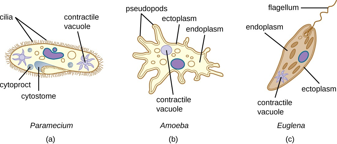
All protozoans have a plasma membrane, or plasmalemma. Many protists have whip-like flagella or hair-like cilia made of microtubules that can be used for locomotion (Figure 4.3). Other protists use cytoplasmic extensions known as pseudopodia (“false feet”) to attach the cell to a surface; they then allow cytoplasm to flow into the extension, thus moving themselves forward.
Protozoans have a variety of unique organelles and sometimes lack organelles found in other cells. Some have contractile vacuoles, organelles that can be used to move water out of the cell for osmotic regulation (salt and water balance) (Figure 4.3). Mitochondria may be absent in parasites or altered to kinetoplastids (modified mitochondria) or hydrogenosomes.
![]()
- What is the sequence of events in reproduction by schizogony and what are the cells produced called?
Taxonomy of Protists
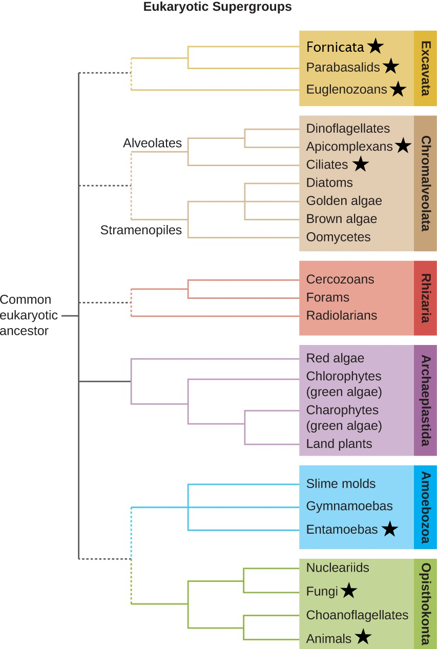
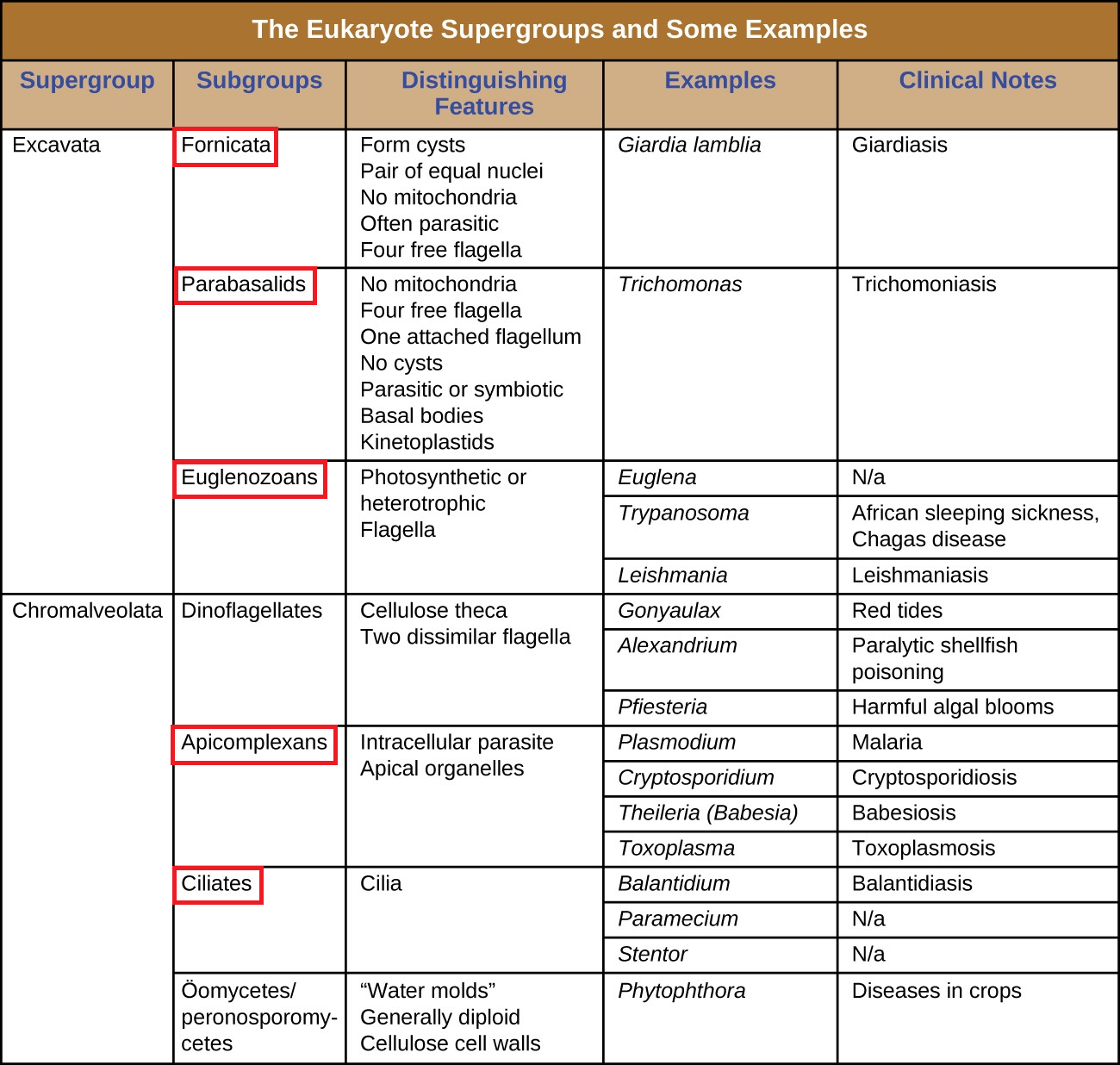
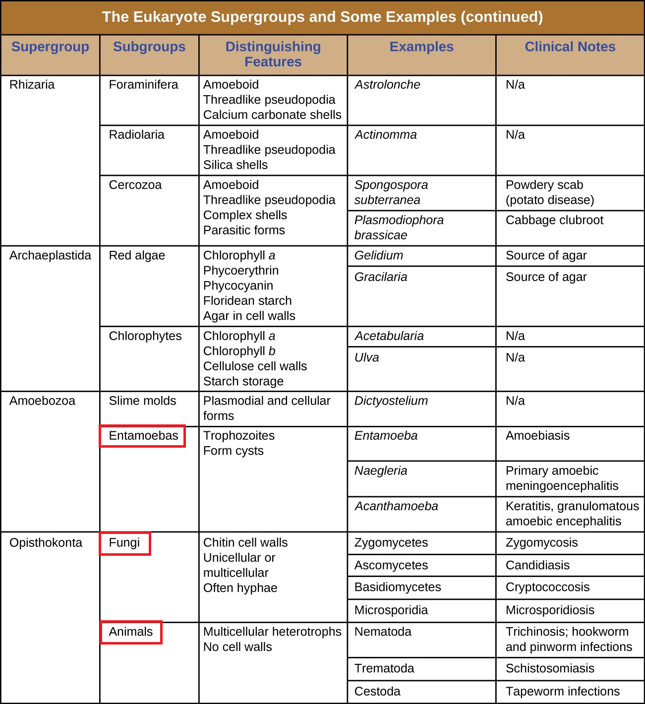
The protists are a polyphyletic group, meaning they lack a shared evolutionary origin. Since the current taxonomy is based on evolutionary history (as determined by biochemistry, morphology, and genetics), protists are scattered across many different taxonomic groups within the domain Eukarya. Eukarya is currently divided into six supergroups that are further divided into subgroups, as illustrated in (Figure 4.4). In this section, we will primarily be concerned with the subgroups Formicata, Parabasalids, Eugoenozoans, Apicomplexan, Ciliates, and Entamoebas, ; since these subgroups include many protozoans of clinical significance. Figure 4.5 and Figure 4.6 summarize the characteristics of each supergroup and subgroup and list representatives of each.
![]()
- Which subgroups contain the clinically significant protists?
Subgroup Entamoeba
The subgroup Entameoba includes protozoans that use amoeboid movement. Actin microfilaments produce pseudopodia, into which the remainder of the protoplasm flows, thereby moving the organism. Members of this subgroup are the primary cause of amoebic dysentery.
Subgroups Apicomplexan and Ciliates
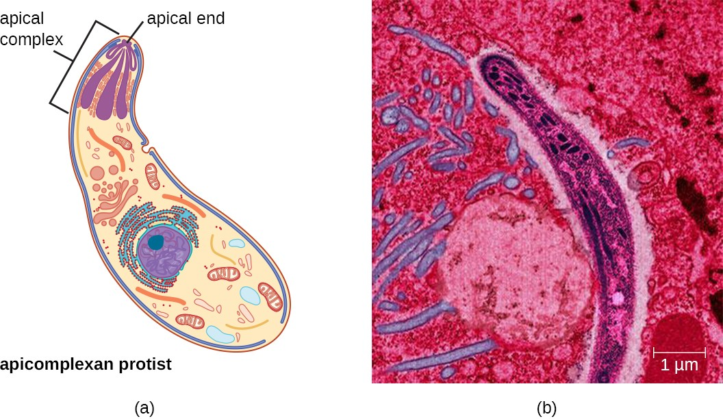
The subgroups Apicomplexan and Ciliates are united by similar origins of its members’ plastids. The apicomplexans are intra- or extracellular parasites that have an apical complex at one end of the cell. The apical complex is a concentration of organelles, vacuoles, and microtubules that allows the parasite to enter host cells (Figure 4.7). Many are capable of infecting a variety of animal cells, from insects to livestock to humans, and their life cycles often depend on transmission between multiple hosts.
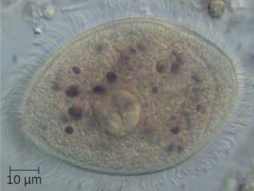
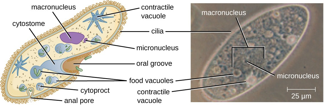
The ciliates (Ciliaphora) are a large, very diverse group characterized by the presence of cilia on their cell surface. Although the cilia may be used for locomotion, they are often used for feeding, as well, and some forms are nonmotile. Balantidium coli (Figure 4.8) is the only parasitic ciliate that affects humans by causing intestinal illness, although it rarely causes serious medical issues except in the immunocompromised (those having a weakened immune system). Perhaps the most familiar ciliate is Paramecium, a motile organism with a clearly visible cytostome and cytoproct that is often studied in biology laboratories (Figure 4.9).
Ciliates are able to reproduce through conjugation, in which two cells attach to each other and exchange DNA, forming cells that are genetically different from each other and from their previous versions.
Link to Learning
Explore the procedures for detecting the presence of an apicomplexan in a public water supply, at this website (https://openstax.org/l/22detpreapicom).
Subgroups Fornicata, Parabasalia, Euglenozoa
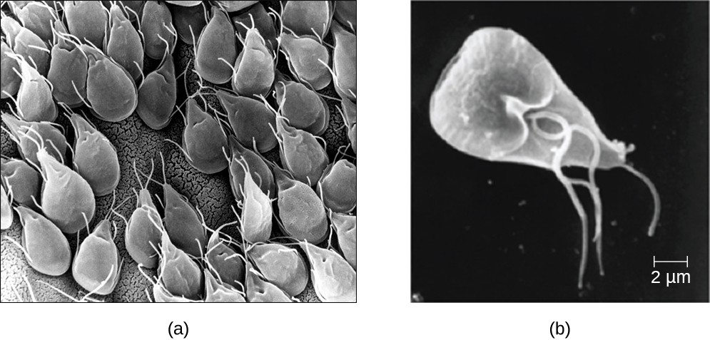
The subgroups Fornicata, Parabasalia, and Euglenozoa includes primitive eukaryotes and many parasites with limited metabolic abilities. These organisms have complex cell shapes and structures, often including a depression on the surface of the cell. The Fornicata lack mitochondria but have flagella. Parabasalia are frequent animal endosymbionts; they live in the guts of animals like termites and cockroaches. They have basal bodies and modified mitochondria (kinetoplastids). They also have a large, complex cell structure with an undulating membrane and often have many flagella. The trichomonads (a subgroup of the Parabasalia) include pathogens such as Trichomonas vaginalis, which causes the human sexually transmitted disease trichomoniasis.
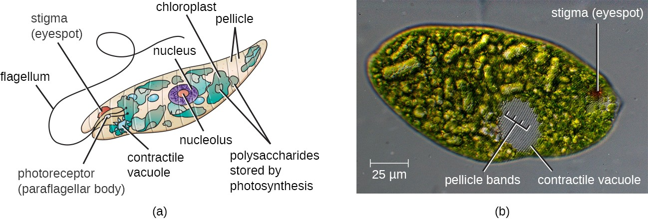
The Euglenozoa are common in the environment and include photosynthetic and nonphotosynthetic species. Their cells have two flagella, a pellicle, a stigma (eyespot) to sense light, and chloroplasts for photosynthesis (Figure 4.10). Members of the genus Euglena are typically not pathogenic, but include the trypanosomes, which are parasitic pathogens that cause African sleeping sickness and American trypanosomiasis (Chagas disease). These tropical diseases are spread by insect bites. The early symptoms include confusion, difficulty sleeping, and lack of coordination. Left untreated, it is fatal.