1.3 Types of Microorganisms
Learning Objectives
- List the various types of microorganisms and describe their defining characteristics
- Give examples of different types of cellular and viral microorganisms and infectious agents
- Describe the similarities and differences between archaea and bacteria
- Provide an overview of the field of microbiology
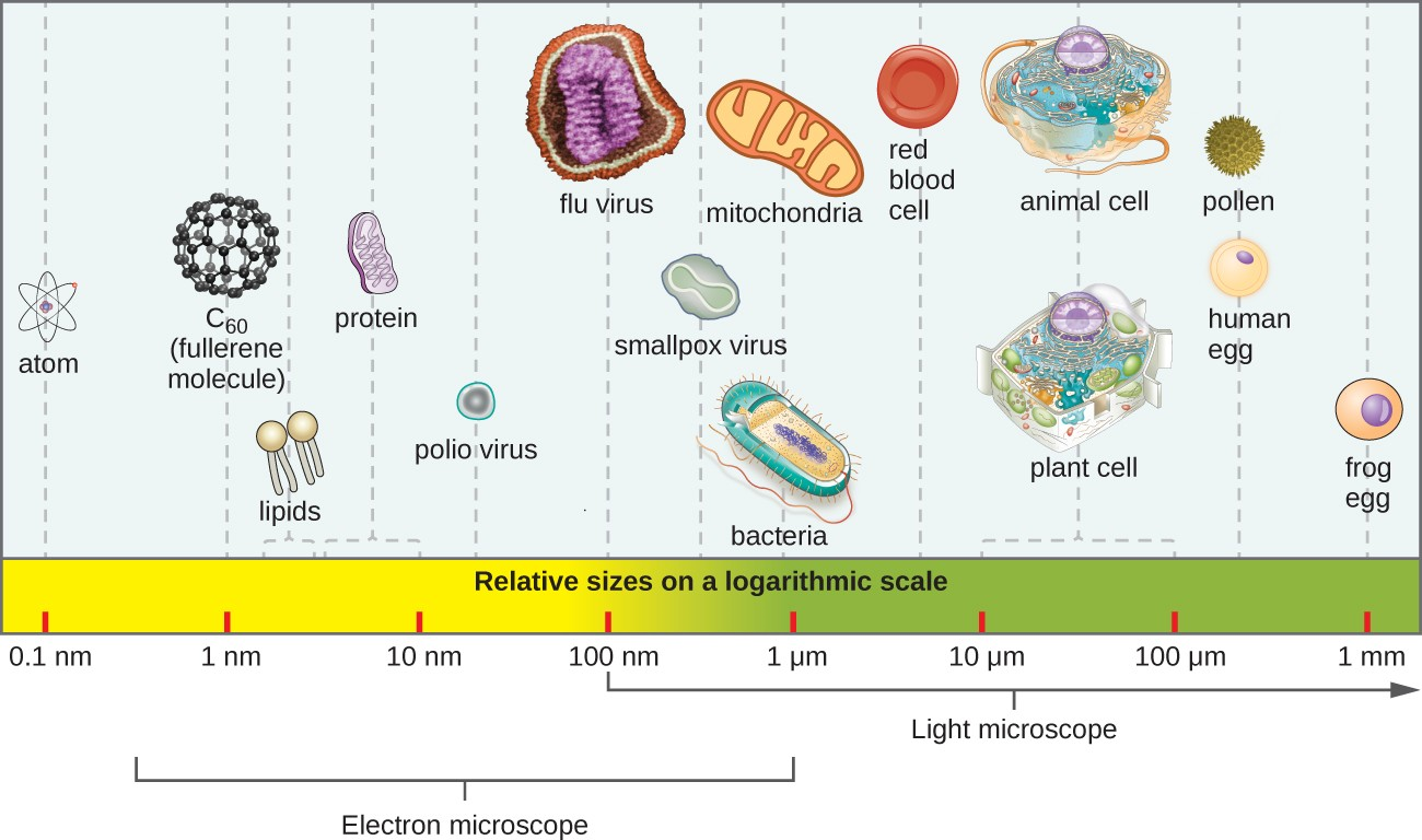
Most microbes are unicellular and small enough that they require artificial magnification to be seen. However, there are some unicellular microbes that are visible to the naked eye, and some multicellular organisms that are microscopic. An object must measure about 100 micrometers (µm) to be visible without a microscope, but most microorganisms are many times smaller than that. For some perspective, consider that a typical animal cell measures roughly 10 µm across but is still microscopic. Bacterial cells are typically about 1 µm, and viruses can be 10 times smaller than bacteria (Figure 1.10). See Table 1.1 for units of length used in microbiology.
|
Metric Unit |
Meaning of Prefix |
Metric Equivalent |
|---|---|---|
|
meter (m) |
— |
1 m = 100 m |
|
decimeter (dm) |
1/10 |
1 dm = 0.1 m = 10−1 m |
|
centimeter (cm) |
1/100 |
1 cm = 0.01 m = 10−2 m |
|
millimeter (mm) |
1/1000 |
1 mm = 0.001 m = 10−3 m |
|
micrometer (μm) |
1/1,000,000 |
1 μm = 0.000001 m = 10−6 m |
|
nanometer (nm) |
1/1,000,000,000 |
1 nm = 0.000000001 m = 10−9 m |
Microorganisms differ from each other not only in size, but also in structure, habitat, metabolism, and many other characteristics. While we typically think of microorganisms as being unicellular, there are also many multicellular organisms that are too small to be seen without a microscope. Some microbes, such as viruses, are even acellular (not composed of cells).
Microorganisms are found in each of the three domains of life: Archaea, Bacteria, and Eukarya. Microbes within the domains Bacteria and Archaea are all prokaryotes (their cells lack a nucleus), whereas microbes in the domain Eukarya are eukaryotes (their cells have a nucleus). Some microorganisms, such as viruses, do not fall within any of the three domains of life. In this section, we will briefly introduce each of the broad groups of microbes. Later chapters will go into greater depth about the diverse species within each group.
Link to Learning
How big is a bacterium or a virus compared to other objects? Check out this interactive website (https://www.openstax.org/l/22relsizes) to get a feel for the scale of different microorganisms.
Prokaryotic Microorganisms
Bacteria are found in nearly every habitat on earth, including within and on humans. Most bacteria are harmless or helpful, but some are pathogens, causing disease in humans and other animals. Bacteria are prokaryotic because their genetic material (DNA) is not housed within a true nucleus. Most bacteria have cell walls that contain peptidoglycan.
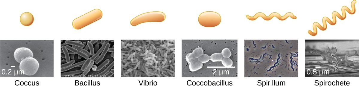
Bacteria are often described in terms of their general shape. Common shapes include spherical (coccus), rod-shaped (bacillus), or curved (spirillum, spirochete, or vibrio). Figure 1.11 shows examples of these shapes.
They have a wide range of metabolic capabilities and can grow in a variety of environments, using different combinations of nutrients. Some bacteria are photosynthetic, such as oxygenic cyanobacteria and anoxygenic green sulfur and green nonsulfur bacteria; these bacteria use energy derived from sunlight, and fix carbon dioxide for growth. Other types of bacteria are nonphotosynthetic, obtaining their energy from organic or inorganic compounds in their environment.
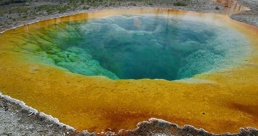
Archaea are also unicellular prokaryotic organisms. Archaea and bacteria have different evolutionary histories, as well as significant differences in genetics, metabolic pathways, and the composition of their cell walls and membranes. Unlike most bacteria, archaeal cell walls do not contain peptidoglycan, but their cell walls are often composed of a similar substance called pseudopeptidoglycan. Like bacteria, archaea are found in nearly every habitat on earth, even extreme environments that are very cold, very hot, very basic, or very acidic (Figure 1.12). Some archaea live in the human body, but none have been shown to be human pathogens.
![]()
- What are the two main types of prokaryotic organisms?
- Name some of the defining characteristics of each type.
Eukaryotic Microorganisms
The domain Eukarya contains all eukaryotes, including uni- or multicellular eukaryotes such as protists, fungi, plants, and animals. The major defining characteristic of eukaryotes is that their cells contain a nucleus.
Protists
Protists are an informal grouping of eukaryotes that are not plants, animals, or fungi. Algae and protozoa are examples of protists.
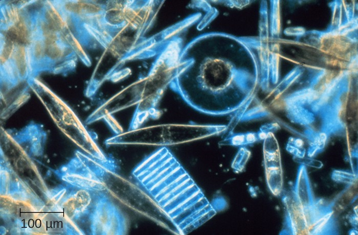
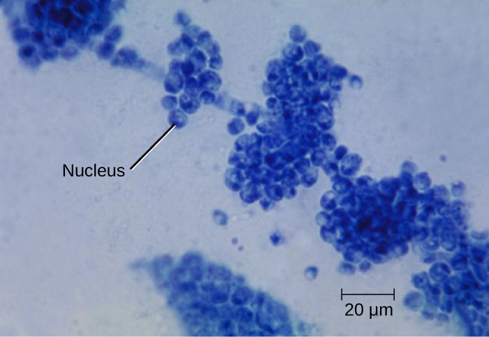
Algae (singular: alga) are protists that can be either unicellular or multicellular and vary widely in size, appearance, and habitat (Figure 1.13). Their cells are surrounded by cell walls made of cellulose, a type of carbohydrate. Algae are photosynthetic organisms that extract energy from the sun and release oxygen and carbohydrates into their environment. Because other organisms can use their waste products for energy, algae are important parts of many ecosystems. Many consumer products contain ingredients derived from algae, such as carrageenan or alginic acid, which are found in some brands of ice cream, salad dressing, beverages, lipstick, and toothpaste. A derivative of algae also plays a prominent role in the microbiology laboratory. Agar, a gel derived from algae, can be mixed with various nutrients and used to grow microorganisms in a Petri dish. Algae are also being developed as a possible source for biofuels.
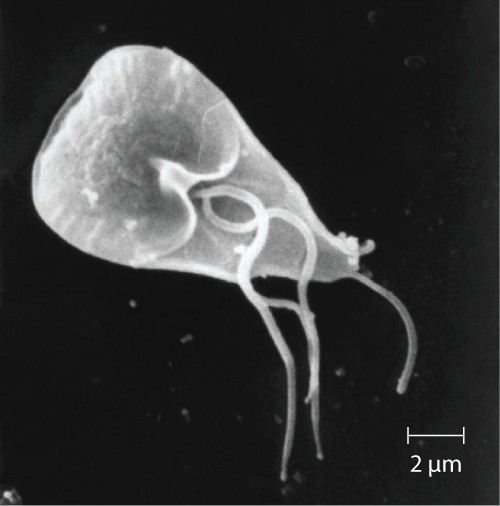
Protozoa (singular: protozoan) are protists that make up the backbone of many food webs by providing nutrients for other organisms. Protozoa are very diverse. Some protozoa move with help from hair-like structures called cilia or whip-like structures called flagella. Others extend part of their cell membrane and cytoplasm to propel themselves forward. These cytoplasmic extensions are called pseudopods (“false feet”). Some protozoa are photosynthetic; others feed on organic material. Some are free-living, whereas others are parasitic, only able to survive by extracting nutrients from a host organism. Most protozoa are harmless, but some are pathogens that can cause disease in animals or humans (Figure 1.14).
Fungi
Fungi (singular: fungus) are also eukaryotes. Some multicellular fungi, such as mushrooms, resemble plants, but they are actually quite different. Fungi are not photosynthetic, and their cell walls are usually made out of chitin rather than cellulose.
Unicellular fungi—yeasts—are included within the study of microbiology. There are more than 1000 known species. Yeasts are found in many different environments, from the deep sea to the human navel. Some yeasts have beneficial uses, such as causing bread to rise and beverages to ferment; but yeasts can also cause food to spoil. Some even cause diseases, such as vaginal yeast infections and oral thrush (Figure 1.15).
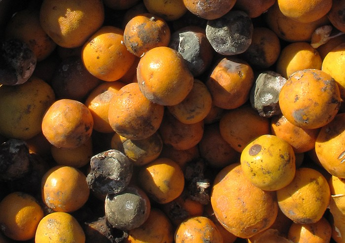
Other fungi of interest to microbiologists are multicellular organisms called molds. Molds are made up of long filaments that form visible colonies (Figure 1.16). Molds are found in many different environments, from soil to rotting food to dank bathroom corners. Molds play a critical role in the decomposition of dead plants and animals. Some molds can cause allergies, and others produce disease-causing metabolites called mycotoxins. Molds have been used to make pharmaceuticals, including penicillin, which is one of the most commonly prescribed antibiotics, and cyclosporine, used to prevent organ rejection following a transplant.
![]()
- Name two types of protists and two types of fungi.
- Name some of the defining characteristics of each type.
Helminths
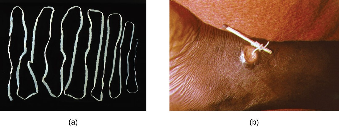
Multicellular parasitic worms called helminths are not technically microorganisms, as most are large enough to see without a microscope. However, these worms fall within the field of microbiology because diseases caused by helminths involve microscopic eggs and larvae. One example of a helminth is the guinea worm, or Dracunculus medinensis, which causes dizziness, vomiting, diarrhea, and painful ulcers on the legs and feet when the worm works its way out of the skin (Figure 1.17). Infection typically occurs after a person drinks water containing water fleas infected by guinea-worm larvae. In the mid-1980s, there were an estimated 3.5 million cases of guinea-worm disease, but the disease has been largely eradicated. In 2014, there were only 126 cases reported, thanks to the coordinated efforts of the World Health Organization (WHO) and other groups committed to improvements in drinking water sanitation.[1][2]
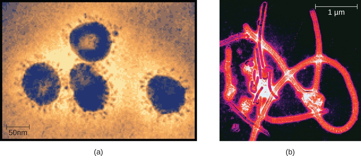
Viruses
Viruses are acellular microorganisms, which means they are not composed of cells. Essentially, a virus consists of proteins and genetic material—either DNA or RNA, but never both—that are inert outside of a host organism. However, by incorporating themselves into a host cell, viruses are able to co-opt the host’s cellular mechanisms to multiply and infect other hosts.
Viruses can infect all types of cells, from human cells to the cells of other microorganisms. In humans, viruses are responsible for numerous diseases, from the common cold to deadly Ebola (Figure 1.18). However, many viruses do not cause disease.
![]()
- Are helminths microorganisms? Explain why or why not.
- How are viruses different from other microorganisms?
Microbiology as a Field of Study
Microbiology is a broad term that encompasses the study of all different types of microorganisms. But in practice, microbiologists tend to specialize in one of several subfields. For example, bacteriology is the study of bacteria; mycology is the study of fungi; protozoology is the study of protozoa; parasitology is the study of helminths and other parasites; and virology is the study of viruses (Figure 1.18). Immunology, the study of the immune system, is often included in the study of microbiology because host–pathogen interactions are central to our understanding of infectious disease processes. Microbiologists can also specialize in certain areas of microbiology, such as clinical microbiology, environmental microbiology, applied microbiology, or food microbiology.
In this textbook, we are primarily concerned with clinical applications of microbiology, but since the various subfields of microbiology are highly interrelated, we will often discuss applications that are not strictly clinical.
