II. Gastrointestinal Tract, Digestive Organs, and Processes
New Terms
Abomasum
Carnivore
Cecum
Colon
Crop
Duodenum
Foregut/Hind-Gut Fermenter
Gizzard
Herbivore
Ileum
Jejunum
Monogastrics
Omasum
Omnivore
Reticulum
Rumen
Villi
Chapter Objective
- To introduce the different organs of the gastrointestinal tract in omnivores and herbivores that are involved in digestion and absorption.
Many animals, such as cows, have multiple, compartmentalized stomachs and are commonly referred to as ruminants. Animals such as pigs, dogs, and chickens have simple noncompartmentalized stomachs and are commonly referred to as nonruminants or monogastrics.
Physiologically, monogastric animals are autoenzymatic digesters (auto = self) in the sense that the enzymes secreted by the animal itself are involved in the digestive processes, whereas ruminant animals are alloenzymatic digesters, in which digestion is accomplished by “others” (allo = others) or microbes residing in the gut. Since the microbes are involved in the “processing” of the ingested feed, instead of “digesting,” fermenting may be the more appropriate term to designate digestive processes in ruminant animals. The site of fermentation varies in alloenzymatic digesters. For example, cattle and sheep are foregut fermenters, while horses and rabbits are hindgut fermenters. Overall, food-producing animals such as pigs and chickens and companion animals (cats, dogs) have a pouch-like, noncompartmentalized stomach, whereas ruminant animals (cows, sheep) have more specialized fermenting chambers. Classification of animals by digestive tract fermentation site is shown in Table 2.1.
| Site of Fermentation | Example |
| Fore-gut | Cattle, sheep, elk, deer |
| Hind-gut | |
| Cecal | Rabbit |
| Colonic | Horse |
| Ceco-colonic | Elephant |
Ruminants
- Multiple compartments
- High storage capacity
- Microbial fermentation
- Microbes provide energy, protein, and B vitamins
Monogastric
- Single compartment
- Limited capacity
- Limited microbial fermentation
- Concentrated feeds and well-balanced diets (e.g., amino acids / vitamins) needed
Monogastric Animals: Gastrointestinal Tract and Digestive Process
The gastrointestinal tract prepares the food (feed) for digestion and absorption. Absorption is the passage of digested nutrients through the gut wall. Digestive processes include mechanical, chemical, and enzymatic processes.
The overall function of the digestive processes is to reduce the feed to a molecular size or increase solubility that allows for absorption and utilization of nutrients from the feed by the cells of the GI tract.
In monogastric animals, digestive processes start with the mouth, tongue, and esophagus and continue through the stomach and small and large intestines. These structures are associated with feed reception, mastication/chewing/swallowing (mouth and tongue), and digestion and absorption (stomach and small and large intestines). The other associated organs involved in the digestive processes are the liver (bile secretion) and pancreas (secretion of several enzymes and hormones).
Features such as the tongue, lips, and/or beak or mouth help in the feed prehension (grabbing) process, and teeth help in chewing and breaking the feed into smaller particles and swallowing. The food is mixed with saliva, which helps in moistening the feed and swallowing. The saliva also contains enzymes such as amylase and lipase (more in newborn animals). The saliva produced by the salivary glands (parotid, submaxillary, sublingual) is added during mastication. Saliva helps in bolus formation and softening of feed, as well as antibacterial action. The enzyme effects of saliva (e.g., amylase, lipase) are minimal due to the short time feed is present in the mouth.
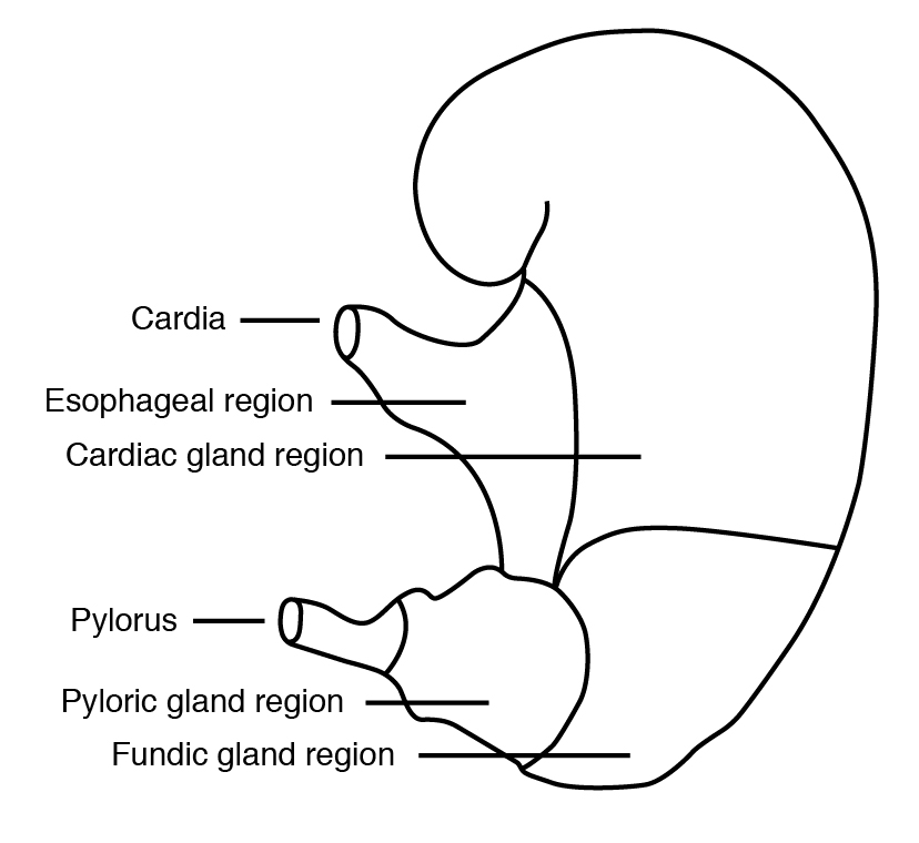
Feed moves down through the esophagus in peristaltic movements through the esophagus into the stomach. There are no glandular secretions in the esophageal region. The stomach is a muscular organ. Functions of the stomach are to serve as a portal or storage of consumed feed and initiate the breakdown of nutrients. The stomach helps in mixing, enzyme secretion, and digestion. The stomach of a monogastric animal includes four functionally distinct zones (Figure 2.1). Adjacent to the esophageal region is the cardiac region, which contains glands that produce mucus. The mucus, a glycoprotein, serves as a protective layer for the stomach wall from acidic secretions and has some antibacterial properties. The fundus gland region and pyloric region are the sites of other secretions such as HCl; gastric secretions, including pepsin; and protein-digesting enzymes. The pH of stomach content varies from one to three due to the acidic action of HCl. The high acidity also provides antibacterial protection to the stomach. The ingested feed exits from the stomach to the duodenum through the pyloric sphincter, which is under hormonal control to not overload the digestive capacity of the small intestine.
Small Intestine
The major site of digestion and absorption in monogastric animals is the small intestine. It is composed of three distinct regions: the duodenum, jejunum, and ileum. The duodenum is the point of entry for secretions such as bile from the gall bladder and secretions from the pancreas.
In order to fit into the small body cavity, the small intestine is highly coiled. In addition to the digestive function, the small intestine is also an immune organ and has special features such as gut-associated lymphoid tissue, Peyer’s patches, and Bursa fabricii (in chickens), serving as part of the body’s immune defense.
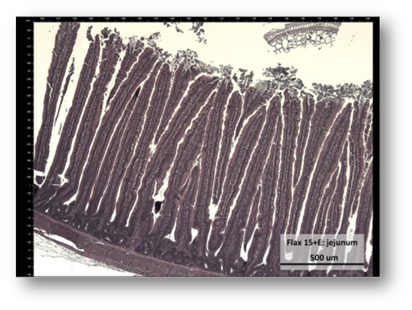
Both digestive and absorptive functions of the small intestine are facilitated by a large surface area. The main increase in surface area is brought about by anatomical features such as villi and microvilli. The villi are small finger-like projections lining the intestinal mucosa and giving it a velvety appearance (Figure 2.2). Each villus is further surrounded by a large number of minute finger-like projections called microvilli. The purpose of these anatomical features is to enhance surface area and thereby absorptive capability (> 60-fold). Each villus contains lymph vessels and capillaries for nutrient transport. The villi are lined with a single layer of cells called enterocytes.
The immature enterocytes are formed in the crypts, and as they mature, they move up the villi, acquiring full complements of the digestive enzymes. As they reach the tip of the villi, they are “worn out” and are extruded into the intestinal lumen. These sloughed-off cells are endogenous (coming from within) in origin and contribute to the metabolic fecal nitrogen. Besides enterocytes, villi also have goblet cells involved in mucus production. The mucus functions as a blanket providing protection from bacterial infection as well as lubrication for digesta.
The large intestine is made up of the cecum, colon, and rectum. The size of the large intestine varies among species based on the type of feed consumed. Hindgut-fermenting animals, such as horses and rabbits, have relatively large ceca. Most digestion and absorption is complete by the time feed residue reaches the colon. The colon functions primarily in water and mineral absorption. Also, some microbial fermentation of plant fiber occurs in the colon. Generally, hindgut fermentation contributes little energy to the animals. However, hindgut fermentation is very important for maintaining gut health and food safety in meat-producing animals.
The liver and pancreas are vital to the digestive process. The liver is the largest gland and is a central organ in nutrient digestion and assimilation. Bile produced from the liver is important for lipid digestion and absorption. The liver plays a role in detoxification of different metabolites as well as storage of many vitamins and minerals. The pancreas produces different enzymes that are needed for the digestion of carbohydrates, proteins, and fats.
Ruminant Animals: Gastrointestinal Tract
The mouth of ruminant animals differs from other mammals in that they have an upper dental pad and not incisor teeth. Feed gathering (prehension) is done by the rough tongue. It is followed by preliminary chewing called mastication and mixing with saliva. Ruminant molars are shaped such that they can chew only on one side at a time. An enormous amount of saliva (about 130 to 150 L/day) is produced in ruminant animals (e.g. dairy cows). Ruminant animals’ saliva is rich in sodium bicarbonate ions and acts as a buffer, aiding in the fermentation process. Ruminant saliva also provides nitrogen (N; urea), phosphorus (P), and sodium (Na), which are utilized by microbes.
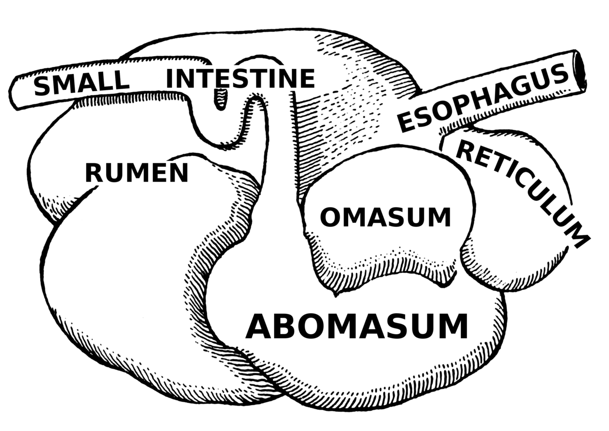
The foregut (stomach) portion of the ruminant animals is divided into four compartments—reticulum, rumen, omasum, and abomasum (Figure 2.3). The reticulum and rumen are partially separated but have different functional purposes.
The rumen is the largest compartment, occupying the left side of the abdominal cavity. The rumen acts as a fermentation vat and is subdivided into sacs by thick muscular boundaries known as pillars. In addition, papillae are also present in the rumen. The rumen has a high population of bacteria, fungi, and protozoa.
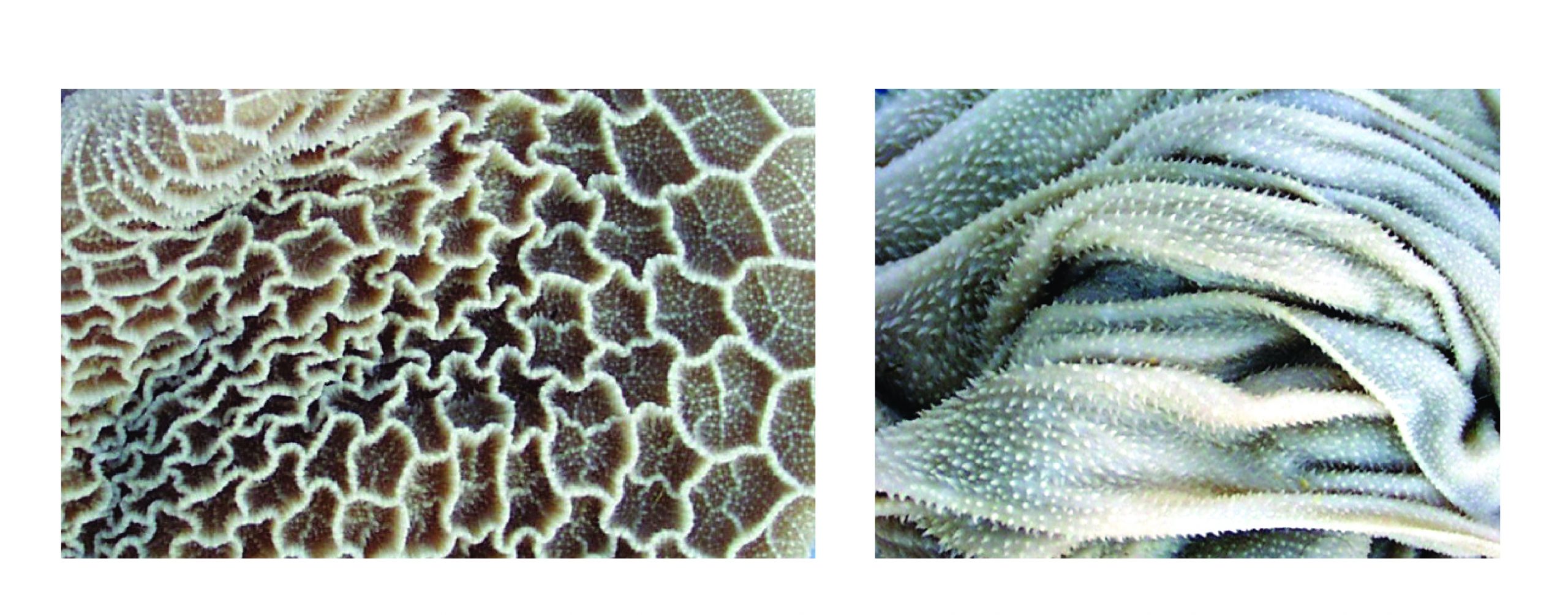
The reticulum is also called the honeycomb because it is lined with a mucous membrane that subdivides the surface into honeycomb-like compartments (Figure 2.4). The reticulum moves ingested feed into the rumen.
The omasum functions as a food filter and aids in reducing particle size (Figure 2.4). The inside of the omasum is thrown into broad longitudinal folds or leaves. Some absorption may occur in the omasum. The omasum regulates flow to the lower gut and absorbs water and some minerals.
The abomasum is the site where the digestive enzymes are first released in ruminants (e.g., pepsin, mucus, HCl). In young animals, the reticulum, rumen, and omasum are relatively underdeveloped, and exposure to feed, the barn, and other environments enables the growth and maturation of the foregut, and this process takes about six to nine months in large ruminants.
Avian Gastrointestinal Tract
Birds such as chickens are also monogastric animals. As with most birds, chickens obtain their food through the use of their beaks, and their oral cavity does not have teeth but has glands that secrete saliva, making feed easy to swallow.
Food then enters the esophagus, which is a flexible tube that connects the mouth with the rest of the digestive tract. The esophagus includes the crop (expansion of the esophagus), which leads to the proventriculus (also known as the true stomach), or the glandular stomach, where digestion primarily begins (Figure 2.5).
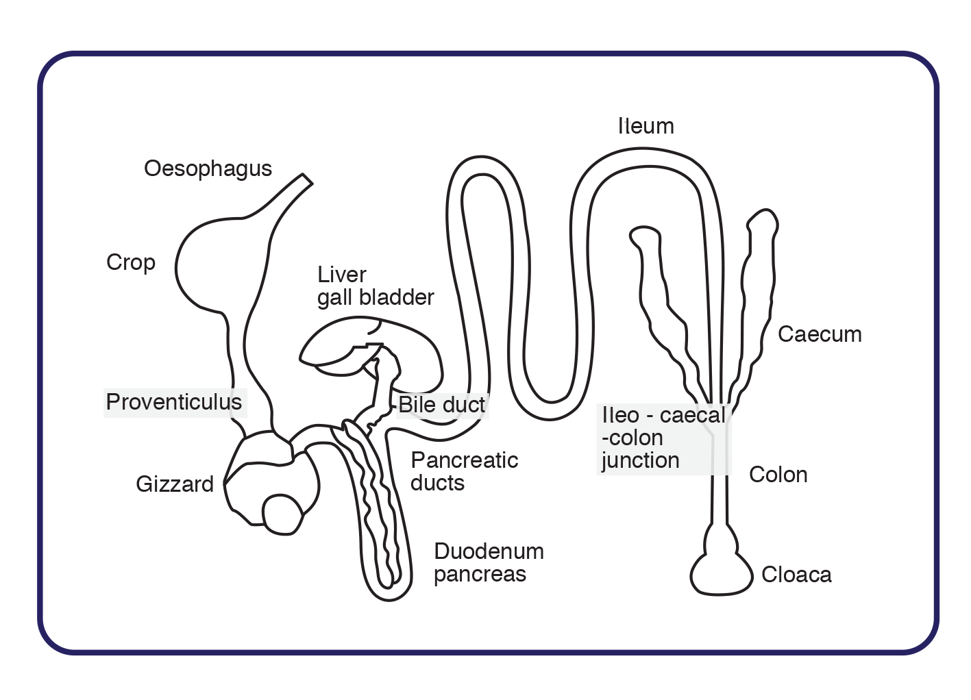
The gizzard is a part of the digestive tract of birds that is often referred to as the mechanical stomach. The gizzard is made up of two sets of strong muscles that act as the bird’s “teeth.” Consumed food passes into the gizzard for grinding and mixing. The gizzard acts only as a mechanical organ; therefore, no digestive aids are secreted and absorption of nutrients does not occur. However, the gizzard is important for mixing ingested feed with water, saliva, hydrochloric acid, and pepsin. The small intestine includes the duodenal loop, which is enclosed by the pancreas. It is also composed of the jejunum and ileum. A remnant attachment of the yolk sac and Meckel’s diverticulum are found at the end of the jejunum. Valves at the end of the ileum control passage to two ceca. After some fermentation, digesta are released into a short large intestine that empties into the cloaca and is eventually excreted through the vent.
Key Points
- Digestive processes include mechanical, chemical, and enzymatic processes.
- Digestive tract variations and specialization are based on diet, and classification is based on the fermentation process.
- Digestion starts in the mouth, where chewing and saliva secretion occur. The functions of saliva include lubrication, buffering, and some enzymatic action.
- The most extensive digestion and absorption in the monogastrics (single stomach) occur in the small intestine.
- The small intestine is divided into the duodenum, jejunum, and ileum. Pancreatic secretions occur in the duodenum.
- The three sections of the large intestine are the cecum, colon, and rectum.
- The four compartments of the ruminant gastrointestinal (GI) tract are the rumen, reticulum, omasum, and abomasum. The abomasum is analogous to the gastric stomach.
- Functions of the reticulum include receiving food, passing food to the omasum, eructation, and rumination.
- The bulk of fermentation occurs in the rumen, where anaerobic bacteria, protozoa, and fungi do their work.
- The omasum regulates flow to the lower gut and absorbs water and some minerals.
- The abomasum is the site where the digestive enzymes are first released in ruminants (although I should say there are trace amounts of salivary lipase in ruminants). Ruminant saliva does not have amylase. This does not matter because microbes secrete a lot of their own amylases.
- Hindgut fermenters are those that use the cecum (or colon) for fermentation of plant fiber. They include birds, pigs, and rabbits. Generally, this is less efficient than foregut fermentation.
- Other accessory organs of the GI tract include the liver, pancreas, and gall bladder.
- In birds, there is an extension of the esophagus called the crop and proventriculus, functioning as the glandular stomach.
- The gizzard is a part of the digestive tract of birds, functioning as the mechanical stomach, where most grinding occurs.
Review Questions
- Compare and contrast the gastrointestinal tract of a swine to that of a cow.
- List the three sections of the small and large intestine.
- The small intestine is the primary site of digestion of several nutrients. What are the special anatomical features of the small intestine helping in this process?
- List the sites of fermentation in foregut fermenting and hindgut fermenting animals.
- List the different sections of the foregut in ruminant animals along with their functions.
