XIII. Vitamins
This chapter provides an introduction and discussion of vitamins that are important in the nutrition of food-producing animals.
New Terms
Antioxidant
Carotenoid
Cholecalciferol
Fat-soluble vitamins
Osteomalacia
Quinones
Retinal
Retinol
Rickets
Tocopherol
Water-soluble vitamins
Chapter Objective
- To introduce and discuss different vitamins of importance in animal health, nutrition, and food quality
What Are Vitamins?
Vitamins are a group of chemically unrelated organic molecules that are needed in minute amounts for different physiological functions. The name “vitamin” originated from the term vital amine and refers to a group of compounds having specific roles in metabolism. Vitamins, although organic compounds, do not provide energy like other macronutrients and are not used for the synthesis of structural compounds. However, they function as enzyme precursors, or coenzymes, in different metabolic processes.
Vitamins
- Do not provide energy like macronutrients
- Function as catalysts in energy-producing reactions
- Make fats more fluid or decreases their melting point when there is an increased amount
Most vitamins need to be provided to the animal through diet, while some of vitamins can be synthesized by the rumen and hindgut microbes or by exposure to sunlight. Deficiency of vitamins in a diet leads to disease conditions, reduced productivity and animal welfare, and reduced immunity in food-producing animals. The dietary requirements of vitamins are very low. In recent years, megadoses of some vitamins (e.g., vitamin E) have been used in animal diets as a means to enhance animal immunity and to improve food quality aspects.
A general classification of vitamins is based on their solubility, as fat- or water-soluble vitamins. The fat-soluble vitamins are vitamin A, vitamin D, vitamin E, and vitamin K. The water-soluble vitamins include members of the B-complex group and vitamin C.
Vitamin Classifications
1. Fat soluble (e.g., vitamin A, vitamin D, vitamin E, vitamin K)
2. Water soluble (e.g., B complex group and vitamin C)
Fat-soluble vitamins
- Vitamins A, D, E, K
- Associated with fat during digestion and absorption
- Storage in liver, adipose tissue, and excess storage can be toxic for some vitamins (e.g., A and D)
- No daily need
- Deficiency is very slow
Water-soluble vitamins
- Total nine, all B vitamins and vitamin C
- Soluble in water and excess excreted through urine
- No storage and less toxic
- Daily requirement (except vitamin B12)
- Serve as cofactor in biochemical reactions
- Deficiency is fast
Fat-Soluble Vitamins
Vitamin A
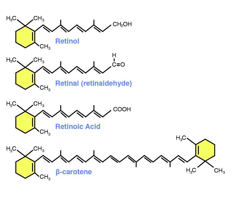
This vitamin was discovered by M. Mori in 1922 as a “fat-soluble factor” present in butter and fish oil, and he named it A. The general term vitamin A includes several related compounds called retinol (alcohol), retinal (aldehyde), and retinoic acid (acid form; figure 13.1). Of these three molecules, retinol is the biologically active form of vitamin A.
Vitamin A is required in the diet of all animals. Vitamin A in the diet can be provided as a vitamin or through its precursor carotenoids present in plants. In animal feeding, most vitamin A is supplied by synthetic sources, which can be produced economically.
Carotenoids are the plant form of or the precursor of vitamin A (figure 13.1). Carotenoids are pigments present in plant cells (> 600 types) that provide the deep orange/yellow color of plant foods such as carrots, sweet potatoes, and pumpkins. There are two forms of carotenoids: carotenes and xanthophylls. Among these, carotenes (especially β-carotenes) have vitamin A activity. The other carotenoids present in plants (xanthophylls) do not have vitamin activity and are involved in providing color pigments. These types of carotenoids are increasingly used in diets for plumage color enrichment (e.g., exotic birds kept in captivity), egg yolk pigmentation, and aquaculture feeds and in the diets of ornamental fish.
Functions
In the body, vitamin A plays a role in several distinct functions, including vision, bone growth, reproduction, and maintenance of epithelial cells, which cover the body surface (e.g., skin) and mucous membranes of body cavities (e.g., respiratory, urogenital, digestive tract).
The role of vitamin A in night vision is well established. In the rods of the retina, retinal combines with a protein called opsin to form rhodopsin (also called visual purple). Rhodopsin is light sensitive and enables the eye to adapt to changes in light intensity. Upon exposure to light, rhodopsin splits into retinal and opsin. The energy that is released is transmitted through the optic nerve leading to vision. However, lack of retinal leads to inefficient rhodopsin recycling making the rod cells insensitive to light changes, eventually leading to night blindness.
Vitamin A is needed for the proliferation and differentiation of cells. Vitamin A is also needed for mucoprotein production, which serves as a barrier and thus protects cells against bacterial invasions. Deficiency of vitamin A can lead to a failure in the differentiation of epithelial cells to mature, mucus-producing cells and to normal epithelial cells being replaced by dysfunctional, stratified, keratinized cells, increasing the susceptibility to infection. Xerophthalmia is a condition in humans and animals that is caused by vitamin A deficiency; it leads to dryness and irritation of the cornea and conjunctiva of the eye and results in cloudiness and infection.
Vitamin A is also needed for normal skeletal and tooth development and reproductive processes. Vitamin A’s role in bone growth is related to its involvement in bone cell (osteoclast and osteoblast) division and maintenance of cell membranes. Vitamin A is also needed for reproductive functions such as spermatogenesis and estrus cycles.
Vitamin A and carotenoids can function as antioxidants thereby protecting cells from oxidative stress and are also involved in modulating cell-mediated and humoral immune responses in animals.
Metabolism
Vitamin A in the diet is digested and absorbed along with fat. In the diet, vitamin A is present as esters, is hydrolyzed by pancreatic lipase, and is incorporated into lipid micelles. Upon reaching the microvilli, they are transferred to mucosal cells, where they are reesterified and are incorporated into the chylomicrons and transported to the lymph for storage in the liver as retinyl esters. The hydrolyzed retinyl esters to free retinol and are complexed with retinol binding proteins and are transported through the blood to the needed tissues.
Carotenoids are split into two within the intestinal mucosal cells to form retinal and are reduced to form retinol. However, a wide variation exists among animals in the bioconversion of carotenoids to retinol. One IU of vitamin A = 0.6 µg of β-carotenes. Some animals, such as cats, cannot convert β-carotene to vitamin A due to the lack of the β-carotene splitting dioxygenase enzyme and need preformed vitamin A from animal sources.
Toxicity
As a fat-soluble vitamin, long-term consumption of vitamin A may lead to toxic symptoms. However, symptoms will vary with species, age, and physiological condition. Skeletal abnormalities and thickening of the skin are reported with hypervitaminosis.
| Functions | Deficiency | Excess |
| Diverse functions such as production of vision pigments, resistance to infectious agents and maintenance of health in many epithelial cells | Impaired growth | Skeletal abnormalities |
| Loss of epithelial integrity | Skin thickening | |
| Reproductive failure | Scaly dermatitis | |
| Swelling and crusting of eyelids |
Vitamin D
Vitamin D includes a group sterol compound that regulates calcium and phosphorus metabolism in the body. Vitamin D is formed by the irradiation of sterols in plants and in the skin of animals and can be called a “sunshine” vitamin. The two major forms of vitamin D are ergocalciferol (vitamin D2, activated plant form) and cholecalciferol (D3, activated animal form; Figure 13.2).

Ergocalciferol (vitamin D2) in plants is formed upon exposure to sunlight after harvest (or injury) and not in living plant cells. Sun-cured forages and hay are good sources of vitamin D in grazing ruminant animals. Animals kept in confinement, as in modern pig and poultry commercial operations, without exposure to sunlight will require vitamin D. The activated animal form of vitamin D3 (cholecalciferol) is the form that is of importance in other omnivores and carnivores. In most animals, vitamin D2 can be converted to vitamin D3. The efficiency of conversion is very low in poultry.
In the body, vitamin D3 is synthesized from cholesterol when it is converted to 7-dehydrocholesterol in the skin upon exposure to ultraviolet irradiation. To become active, it is transported from the skin to the liver, where it is hydroxylated to form 25-hydroxycholecalciferol. This compound is transported through the blood to the kidneys, where it is further hydroxylated to form 1,25-hydroxycholecalciferol, also called calcitriol, which is the most metabolically active form of vitamin D.
Functions
Because vitamin D is produced in the body and due to its regulatory functions in calcium and phosphorus homeostasis, it is also considered as a hormone. In addition to this, other parts of the body (gastrointestinal tract, kidneys, bones) and parathyroid hormones work in conjunction with vitamin D in blood calcium homeostasis and bone calcification. Normal blood calcium levels are achieved by adjusting the dietary absorption of calcium from the gastrointestinal tract and by the release of calcium from the bone. Calcium-binding proteins are needed for proper absorption of calcium and phosphorus from the gut. In the gastrointestinal tract, vitamin D stimulates the synthesis of calcium-binding proteins enabling calcium and phosphorus absorption from the diet. Under a condition of hypocalcemia (low blood Ca level), parathyroid hormones (PTH) stimulate Ca absorption (from gut) and resorption (from bone and kidney tubules) indirectly by stimulating the production of vitamin D.
Vitamin D also affects normal bone growth and calcification by acting with PTH to mobilize Ca from bone and by causing an increase in P resorption in the kidneys. Altogether, vitamin D works along with the intestines, bones, and kidneys to maintain an optimal level of blood Ca and P that is needed for normal bone mineralization.
A deficiency of vitamin D leads to impaired bone mineralization and abnormal skeletal development and results in a condition called rickets in young animals and osteomalacia in growing animals. In each of these instances, inadequate bone calcification leads to lameness, crooked legs, and spontaneous fracture of long bones. Vitamin D deficiency can be prevented by exposure to sunlight for a few minutes, although skin pigmentation affects the amount of sunlight required. White-skinned animals require less sunlight than dark-skinned animals. Similarly, animals such as llamas have higher requirements due to the nature of their high-elevation habitat and exposure to solar radiation. Vitamin D is also used for treating milk fever (discussed in detail in chapter 15) in dairy cows.
Among food sources, egg yolks and organ meats are good sources that could be used in pet animal feeding. Other concentrated sources include cod liver oil. In food animal feeding, vitamin D (cholecalciferol) is included as a Vitamin D supplement.
| Functions | Deficiency | Excess |
| A steroid hormone. | Rickets (young animals) | Abnormal deposition of Ca in soft tissues (kidney, aorta, lungs). |
| Synthesized in the skin when exposed to sunlight | Osteomalacia (growing animals) | |
| Regulates blood Ca level | ||
| Facilitate absorption of calcium from the intestine, and thereby assist in maintaining calcium homeostasis |
Vitamin E
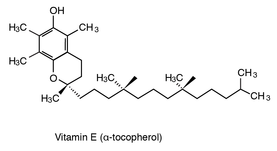
Vitamin E is a term that is used to describe a group of chemically related compounds called tocopherols and tocotrienols. Among the different isomers, α-tocopherol is the most active biological form of vitamin E and is the one that is added to animal diets (Figure 13.3). Other isomers with less biological effects include β-, γ-, δ-tocopherol and α-, β-, γ-, δ tocotrienols. Most commercially available vitamin E is DL-α-tocopheryl acetate. One IU of vitamin E is defined as 1 mg of all-rac-α-tocopherol acetate. The only stereoisomer of α-tocopherol found in nature is RRR-α-tocopherol, which is the most biologically effective form of vitamin E in animals.
Functions
The function of vitamin E in the body is to serve as a biological chain-breaking antioxidant and to protect cells and tissues from oxidative damage induced by free radicals and other lipid oxidation products. Vitamin E prevents the oxidation of lipids by serving as a free radical scavenger and donates electrons from the hydroxyl group of the molecule (antioxidant effect; Figure 13.3). Lipid peroxidation causes damage to unsaturated lipids in cell membranes resulting in the disruption of the structural membrane and cell integrity.
In prepared feeds, the formation of such peroxidized compounds can cause a reduction in palatability, rancidity, and destruction of nutrients and can also affect animal health while reducing organoleptic and sensory quality of the food produced. In addition to lipids and oxidative stress, vitamin E can protect other nutrients such as proteins and vitamin A. Due to these roles, the level of vitamin E in a diet depends on the level of polyunsaturated fatty acids, degree of peroxidative damage, and other external stressors.
Vitamin E also has a sparing action on the mineral selenium, which is a cofactor for the enzyme glutathione peroxidase, which functions to reduce lipid peroxides. The inactivation of lipid peroxides protects the cell membrane from further damage. By preventing the oxidation of the cell membrane polyunsaturated fatty acids, vitamin E spares selenium.
Among food sources, plant oils and egg yolks (depending on hen diet) are good sources of vitamin E. Since vitamin E is highly prone to destruction, proper storage of prepared feed (away from heat and light) is necessary to prevent oxidative changes to fat and to maintain vitamin E levels.
Deficiency
Vitamin E deficiency can produce white muscle disease, exudative diathesis, and encephalomalacia. White muscle disease is caused by the degeneration of skeletal and heart muscle fiber, which leads to rapid death due to heart failure. Exudative diathesis in chickens is caused by leaky capillaries in the breast muscle. Treatment with either vitamin E or selenium will be successful in both cases. However, encephalomalacia (crazy chick disease) can only respond to vitamin E treatment.
Toxicity
Vitamin E is the least toxic of the fat-soluble vitamins and high levels are added in the diets of animals (beef cow, poultry) to enhance food nutritional and aesthetic value and lipid stability.
| Functions | Deficiency | Excess |
| Free radical scavenger | White muscle disease | Non toxic |
| Antioxidant function | Crazy chick disease | High levels are added in animal diets to enhance lipid stability in omega-3 fatty-acid rich foods, increase visual aspects in red meat, and to reduce heat stress and enhance immune function in poultry. |
| Affects immune response | Reduction in feed and food lipid quality and rancidity | |
| Reproductive failure |
Vitamin K
Vitamin K includes a group of compounds called the quinones. Vitamin K1 is found in green plants (phylloquinones) and vitamin K2 (menaquinones) is synthesized by hindgut bacteria. Vitamin K’s are absorbed readily with fat in the gastrointestinal (GI) tract. The liver converts vitamin K1 and K3 to K2 before it is used. The metabolically active form of vitamin K is menaquinones. Menadione (vitamin K3, synthetic form) is the most common version of vitamin K that is included in animal diets.
Functions
Vitamin K is needed for the synthesis of prothrombin, a blood-clotting protein. The blood-clotting process needs several proteins such as thromboplastin, prothrombin, fibrinogen, and fibrin. The enzymes needed for these processes are vitamin K dependent, and hence deficiency of vitamin K leads to failure in fibrin clot formation, hemorrhages, and/or prolonged bleeding time. Often, subcutaneous hemorrhages appear over the body surface, giving a blotchy, bluish appearance to the skin. This can also be of economic importance in food-producing animals leading to a reduction in carcass quality or condemnation.
Gastrointestinal bacterial can provide the needed vitamin K to most animals either through absorption from the hindgut or through coprophagy (eating feces). However, animal husbandry practices, such as confinement housing or raising animals in cages or pens with wire floors, or antibiotic therapy can limit the availability of vitamin K in the animal diet.
Certain coccidiostats containing sulfa drugs can cause vitamin K deficiency as sulfa drugs are an antagonist of vitamin K. Mold growing on weather-damaged sweet clover hay or silage contains dicoumarol, which is very similar to vitamin K in structure. Dicoumarol is a competitive inhibitor of vitamin K. Animals that consume moldy sweet clover hay or silage develop a vitamin K deficiency, leading to internal hemorrhaging and death in calves. Another antagonist of vitamin K is Warfarin, a rat poison causing anticoagulation. It is also a competitive inhibitor of vitamin K. Vitamin K is routinely administered in rodenticide poisoning in pets because the active ingredient (Warfarin) in these rodenticides are anticoagulants, causing bleeding and hemorrhaging.
Phylloquinone and menaquinone derivatives are nontoxic even at higher levels. However, menadione given in prolonged high doses produces anemia and other abnormalities in animals.
| Function | Deficiency | Excess |
| Serves as cofactor in carboxylation reactions of the activation of proteins necessary for blood clotting. | Vitamin K deficiency leads to prolonged blood clotting time and hemorrhaging. Deficiency is rare because vitamin K is synthesized by microbes in the hindgut. Antibiotics and other vitamin K inhibitors in the diet can cause deficiency | Non toxic |
***
Key Points
- Fat-soluble vitamins include vitamins A, D, E, and K.
- They are digested and absorbed along with dietary fat and can be found in the micelle in the gastrointestinal (GI) tract after digestion, and once absorbed into the body, they are transported by chylomicron to various tissues. They are stored in the liver and adipose tissue.
- Vitamin A was discovered by M. Mori in 1922 as a fat-soluble factor.
- Retinol is the chemical name of vitamin A. Retinal and retinoic acid are related compounds. β-carotene, a plant pigment, is the precursor of vitamin A. Vitamin A has two distinct functions: participation in the visual purple cycle in the retina and maintenance of epithelial cells. The former requires retinal, and the latter requires retinoic acid.
- β-carotene can split into two units of retinol in the intestinal mucosa cells; however, this conversion is not efficient. Some animals, such as cats, cannot absorb β-carotene and process it in the liver. Vitamin A activity depends on its double bonds, which are quite labile. An antioxidant is required to protect vitamin A.
- Night blindness, dryness of eyes, diarrhea, kidney stones, and abortion are some typical vitamin A deficiency symptoms. Excessive vitamin A intake will eventually lead to toxicity. Symptoms include dermatitis, skin thickening, and weight loss. Each international unit represents 0.3 µg retinol. The requirement is a 2,000 IU/kg diet.
- Vitamin D is really a hormone. It can be synthesized in the animal body from cholesterol. The whole process involves three different tissues: skin, liver, and kidney; UV light is required.
- Vitamin D, a fat-soluble factor in cod liver oil, is known to cure rickets in humans. Its function is to modulate calcium metabolism in the animal body. Vitamin D increases blood Ca++ concentration by increasing Ca++ absorption from the GI tract and resorption from bone.
- Vitamin D deficiency leads to calcium deficiency. Therefore, deficiency symptoms are the same as those of calcium deficiency, such as abnormal skeletal development.
- Overdose of vitamin D will cause toxicity, which can lead to calcification of soft tissues such as the lungs, aorta, and heart.
- Vitamin E comprises a group of compounds called tocopherols and tocotrienols.
- Tocopherols contain a group of compounds with varying amounts of vitamin E activity; α-tocopherol is the most active one.
- Vitamin E has two functions: avoiding oxidation and promoting normal reproduction in rats. All cell membranes are made of lipid bilayers containing many polyunsaturated fatty acids (PUFAs). Vitamin E serves as an antioxidant to protect the integrity of cell membranes.
- Vitamin E deficiency problems include white muscle disease, exudative diathesis, and encephalomalacia. All these problems occur in young, growing animals. White muscle disease is caused by degeneration of skeletal and heart muscle fiber, which leads to rapid death due to heart failure. Exudative diathesis in chickens is caused by leaky capillaries in the breast muscle. Treatment with either vitamin E or selenium will be successful in both cases. However, encephalomalacia (crazy chick disease) can only respond to vitamin E treatment.
- High levels of PUFA and vitamin A in a diet increase the requirement for vitamin E. It turns out that vitamin E prevents the formation of lipid peroxide from PUFAs, and Se, a cofactor in the enzyme of glutathione peroxidase, is required to remove lipid peroxide.
- Phylloquinone (K1), menaquinone (K2), and menadione (K3) are the three forms of vitamin K. The only known function of vitamin K is as a cofactor in carboxylation reactions of the activation proteins necessary for blood clotting.
- Vitamin K is absorbed readily with fat in the GI tract. The liver converts vitamin K1 and K3 to K2 before it is used. Mold growing on sweet clover contains dicoumarol, which is very similar to vitamin K in structure. Animals that consume moldy sweet clovers develop a vitamin K deficiency. Dicoumarol is a competitive inhibitor of vitamin K.
- Another antagonist to vitamin K is Warfarin, a rat poison. It is also a competitive inhibitor of vitamin K. Vitamin K deficiency leads to prolonged blood-clotting time and hemorrhaging. Deficiency is rare because vitamin K is synthesized by microbes in the hindgut. No toxicity is observed. The requirement of vitamin K (menadione) is a 0.5 mg/kg diet.
Review Questions
- Why can an animal grow well with little vitamin A in its diet but needs to have vitamin B in its diet everyday?
- Name one major deficiency problem resulting from a dietary lack of vitamin A and vitamin K.
- Cows eating sun-cured hay or forage will get this form of vitamin D.
- Ergocalciferol
- Cholecalciferol
- 7-dehydrocholesterol
- Cholesterol
- Exposing your lovebirds to sunshine will help in getting this vitamin.
- Vitamin A
- Vitamin D
- Vitamin B
- Vitamin C
- What are the functions of vitamin E?
- What is coprophagy? Why is it important to animals in terms of vitamin K availability?
- Why is moldy sweet clover toxic to animals?
- Vitamin D is ____.
- An organic compound
- A hormone
- A cholesterol-derived compound
- All of the above
- Drinking milk will give this form of vitamin D.
- Ergocalciferol
- Cholecalciferol
- 7-dehydrocholesterol
- Cholesterol
- The back fat of pigs will have these vitamins in stored form
- Vitamin A
- Vitamin D
- Vitamin B
- Both a and b
