1 Introduction to Microbiology
Welcome to the wonderful world of microbiology! Yay! So. What is microbiology? If we break the word down it translates to “the study of small life,” where the small life refers to microorganisms or microbes. But who are the microbes? And how small are they?
Generally microbes can be divided into two categories: the cellular microbes (or organisms) and the acellular microbes (or agents). In the cellular camp we have the bacteria, the archaea, the fungi, and the protists (a bit of a grab bag composed of algae, protozoa, slime molds, and water molds). Cellular microbes can be either unicellular, where one cell is the entire organism, or multicellular, where hundreds, thousands or even billions of cells can make up the entire organism. In the acellular camp we have the viruses and other infectious agents, such as prions and viroids.
In this textbook the focus will be on the bacteria and archaea (traditionally known as the “prokaryotes,”) and the viruses and other acellular agents.
Characteristics of Microbes
Obviously microbes are small. The traditional definition describes microbes as organisms or agents that are invisible to the naked eye, indicating that one needs assistance in order to see them. That assistance is typically in the form of a microscope of some type. The only problem with that definition is that there are microbes that you can see without a microscope. Not well, but you can see them. It would be easy to dismiss these organisms as non-microbes, but in all other respects they look/act/perform like other well-studied microbes (who follow the size restriction).
So, the traditional definition is modified to describe microbes as fairly simple agents/organisms that are not highly differentiated, meaning even the multicellular microbes are composed of cells that can act independently– there is no set division of labor. If you take a giant fungus and chop half the cells off, the remaining cells will continue to function unimpeded. Versus if you chopped half my cells off, well, that would be a problem. Multicellular microbes, even if composed of billions of cells, are relatively simple in design, usually composed of branching filaments.
It is also acknowledged that research in the field of microbiology will require certain common techniques, largely related to the size of the quarry. Because microbes are so small and there are so many around, it is important to be able to isolate the one type that you are interested in. This involves methods of sterilization, to prevent unwanted contamination, and observation, to confirm that you have fully isolated the microbe that you want to study.
Microbe Size
Since size is a bit of theme in microbiology, let us talk about actual measurements. How small is small? The cellular microbes are typically measured in micrometers (µm). A typical bacterial cell (let us say E. coli) is about 1 µm wide by 4 µm long. A typical protozoal cell (let us say Paramecium) is about 25 µm wide by 100 µm long. There are 1000 µm in every millimeter, so that shows why it is difficult to see most microbes without assistance. (An exception would be a multicellular microbe, such as a fungus. If you get enough cells together in one place, you can definitely see them without a microscope!)
When we talk about the acellular microbes we have to use an entirely different scale. A typical virus (let us say influenza virus) has a diameter of about 100 nanometers (nm). There are 1000 nanometers in every micrometer, so that shows why you need a more powerful microscope to see a virus. If a typical bacterium (let us pick on E.coli again) were inflated to be the size of the Statue of Liberty, a typical virus (again, influenza virus works) would be the size of an adult human, if we keep the correct proportions.
http://learn.genetics.utah.edu/content/cells/scale/
The Discovery of Microbes
The small size of microbes definitely hindered their discovery. It is hard to get people to believe that their skin is covered with billions of small creatures, if you cannot show it to them. “Seeing is believing,” that is what I always say. Or someone says that.
In microbiology, there are two people that are given the credit for the discovery of microbes. Or at least providing the proof of their discovery, both around the same time period:
Robert Hooke (1635-1703)
Robert Hooke was a scientist who used a compound microscope, or microscope with two lenses in tandem, to observe many different objects. He made detailed drawings of his observations, publishing them in the scientific literature of the day, and is credited with publishing the first drawings of microorganisms. In 1665 he published a book by the name of Micrographia, with drawing of microbes such as fungi, as well as other organisms and cell structures. His microscopes were restricted in their resolution, or clarity, which appeared to limit what microbes he was able to observe.
Antony van Leeuwenhoek (1632-1723)
Antony van Leeuwenhoek was a Dutch cloth merchant, who also happened to dabble in microscopes. He constructed a simple microscope (which has a single lens), where the lens was held between two silver plates. Apparently he relished viewing microbes from many different sample types – pond water, fecal material, teeth scrapings, etc. He made detailed drawings and notes about his observations and discoveries, sending them off to the Royal Society of London, the scientific organization of that time. This invaluable record clearly indicates that he saw both bacteria and a wide variety of protists. Some microbiologists refer to van Leeuwenhoek as the “Father of Microbiology,” because of his contributions to the field.
Microbial Groups
Classification of organisms, or the determination of how to group them, continually changes as we acquire new information and new tools of assessing the characteristics of an organism. Currently all organisms are grouped into one of three categories or domains: Bacteria, Archaea, and Eukarya. The Three Domain Classification, first proposed by Carl Woese in the 1970s, is based on ribosomal RNA (rRNA) sequences and widely accepted by scientists today as the most accurate current portrayal of organism relatedness.
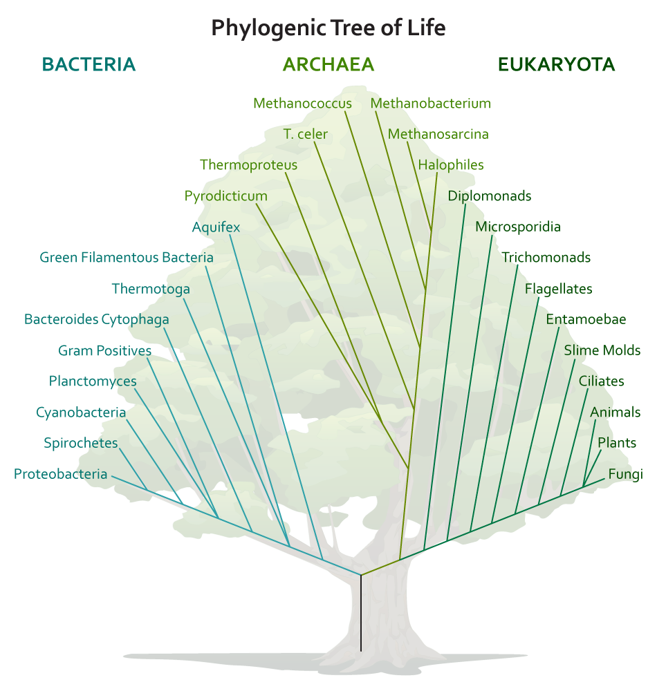
Bacteria
The Bacteria domain contains some of the best known microbial examples (E. coli, anyone?). Most of the members are unicellular (but not all!), most members lack a nucleus or any other organelle, most members have a cell wall with a particular substance known as peptidoglycan (not found anywhere else but in bacteria!), they have 70S ribosomes, and humans are intimately familiar with many members, since they are common in soil, water, our foods, and our own bodies. All Bacteria are considered microbes.
Archaea
Archaea is a relatively new domain, since these organisms used to be grouped with the bacteria. There are some obvious similarities, since they are mostly (but not all!) unicellular, cells lack a nucleus or any other organelle, they have 70S ribosomes, and all Archaea are microbes. But they have completely different cell walls that can vary markedly in composition (but notably lack peptidoglycan and might have pseudomurien instead). In addition, their rRNA sequences have shown that they are not closely related to Bacteria at all.
Eukarya
The Eukarya Domain includes many non-microbes, such as animals and plants, but there are numerous microbial examples as well, such as fungi, protists, slime molds, and water molds. The eukaryotic cell type has a nucleus, as well as many organelles, such as mitochondria or an endoplasmic reticulum. They have 80S ribosomes and are commonly found as unicellular or multicellular.
Viruses
Viruses are not part of the Three Domain Classification, since they lack ribosomes and therefore lack rRNA sequences for comparison. They are classified separately, using characteristics specific to viruses. Viruses are typically described as “obligate intracellular parasites,” a reference to their strict requirement for a host cell in order to replicate or increase in number. These acellular entities are often agents of disease, a result of their cell invasion.
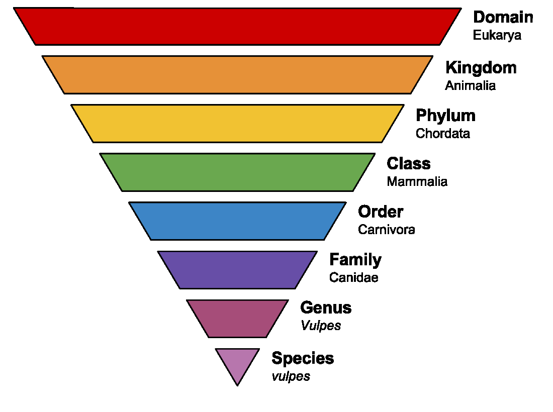
Taxonomic Ranks
Taxonomic ranks are a way for scientists to organize information about organisms, by determining relatedness. Domains are the largest grouping used, followed by numerous smaller groupings, where each smaller grouping consists of organisms that share specific features in common. Each level becomes more and more restrictive as to whom can be a member. Eventually we get down to genus and species, the groupings used for formation of a scientific name. This is the binomial nomenclature devised by Carl Linnaeus in the 1750s.
Binomial Nomenclature
When referring to the actual scientific name assigned to an organism, it is important to follow convention, so it is clear to everyone that you are referring to the scientific name. There are rules in science (just like in English class, where you would never refer to “mr. robert louis stevenson,” or at least not without expecting to get your paper back with red all over it).
A scientific name is composed of a genus and a species, where the genus is a generic name and the species is specific. The species name, once assigned, is permanent for the organism, while the genus can change if new information becomes available. For example, the bacterium previously known as Streptococcus faecalis is now Enterococcus faecalis because sequencing information indicates that it is more closely related to the members of the Enterococcus genus. It is important to note that it is inappropriate to refer to an organism by the species alone (i.e. you should never refer to E. coli as “coli” alone. Other bacteria can have the species “coli” as well.)
Now for the rules: The genus is always capitalized. The species is always lowercase. And both the genus and the species are italicized (common if typewritten) or underlined (common if handwritten). The genus may be shortened to its starting letter, but only if the name has been referred to in the text in its entirety at least once first (the exception to this is E. coli, due to its commonality, where hardly anyone spells out the Escherichia genus anymore).
Key Words
microbiology, microorganisms, microbes, unicellular, multicellular, differentiation, sterilization, observation, micrometers (µm), nanometers (nm), Robert Hooke, compound microscope, Antony van Leeuwenhoek, simple microscope, Royal Society of London, Father of Microbiology, Three Domain Classification, ribosomal RNA (rRNA), Bacteria, Archaea, Eukarya, obligate intracellular parasites, taxonomic ranks, genus, species, binomial nomenclature
Study Questions
- Who are the members of the microbial world?
- What is the complete definition of microbiology? What characteristics are relevant?
- What size are different groups of microbes?
- What were the contributions of Hooke and Van Leeuwenhoek to the field of microbiology? How did they make these contributions?
- What is the basis for Woese’s classification and what are the three domains?
- What are the basic characteristics of members of the three domains? Where do microbes fit in?
- What are the basic characteristics of viruses? Why are they not classified in one of the three domains?
- What are taxonomic ranks? What is the system of binomial nomenclature? What are the basic rules? How are bacteria named? What is a genus and species? Be able to write a bacterial name correctly.
