15.4 Equilibrium
Learning Objectives
By the end of this section, you will be able to:
- Describe the means of mechanoreception for hearing
The Vestibular System (Equilibrium)
Along with audition, the inner ear is responsible for encoding information about equilibrium, the sense of balance. A similar mechanoreceptor—a hair cell with stereocilia—senses head position, head movement, and whether our bodies are in motion. These cells are located within the vestibule of the inner ear. Head position is sensed by the utricle and saccule, whereas head movement is sensed by the semicircular canals. The neural signals generated in the vestibular ganglion are transmitted through the vestibulocochlear nerve to the brain stem and cerebellum.
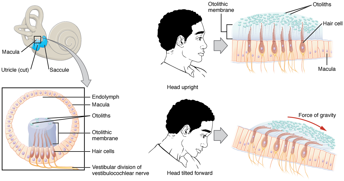
The utricle and saccule are both largely composed of macula tissue (plural = maculae). The macula is composed of hair cells surrounded by support cells. The stereocilia of the hair cells extend into a viscous gel called the otolithic membrane (Figure 15.4.1). On top of the otolithic membrane is a layer of calcium carbonate crystals, called otoliths. The otoliths essentially make the otolithic membrane top-heavy. The otolithic membrane moves separately from the macula in response to head movements. Tilting the head causes the otolithic membrane to slide over the macula in the direction of gravity. The moving otolithic membrane, in turn, bends the sterocilia, causing some hair cells to depolarize as others hyperpolarize. The exact position of the head is interpreted by the brain based on the pattern of hair-cell depolarization.
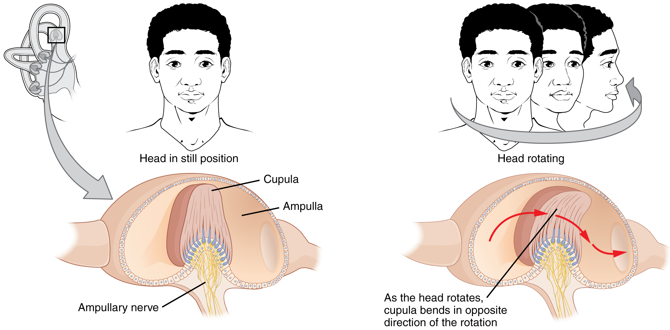
The semicircular canals are three ring-like extensions of the vestibule. One is oriented in the horizontal plane, whereas the other two are oriented in the vertical plane. The anterior and posterior vertical canals are oriented at approximately 45 degrees relative to the sagittal plane (Figure 15.4.2). The base of each semicircular canal, where it meets with the vestibule, connects to an enlarged region known as the ampulla. The ampulla contains the hair cells that respond to rotational movement, such as turning the head while saying “no.” The stereocilia of these hair cells extend into the cupula, a membrane that attaches to the top of the ampulla. As the head rotates in a plane parallel to the semicircular canal, the fluid lags, deflecting the cupula in the direction opposite to the head movement. The semicircular canals contain several ampullae, with some oriented horizontally and others oriented vertically. By comparing the relative movements of both the horizontal and vertical ampullae, the vestibular system can detect the direction of most head movements within three-dimensional (3-D) space.
Central Processing of Vestibular Information
Balance is coordinated through the vestibular system, the nerves of which are composed of axons from the vestibular ganglion that carries information from the utricle, saccule, and semicircular canals. The system contributes to controlling head and neck movements in response to vestibular signals. An important function of the vestibular system is coordinating eye and head movements to maintain visual attention. Most of the axons terminate in the vestibular nuclei of the medulla. Some axons project from the vestibular ganglion directly to the cerebellum, with no intervening synapse in the vestibular nuclei. The cerebellum is primarily responsible for initiating movements on the basis of equilibrium information.
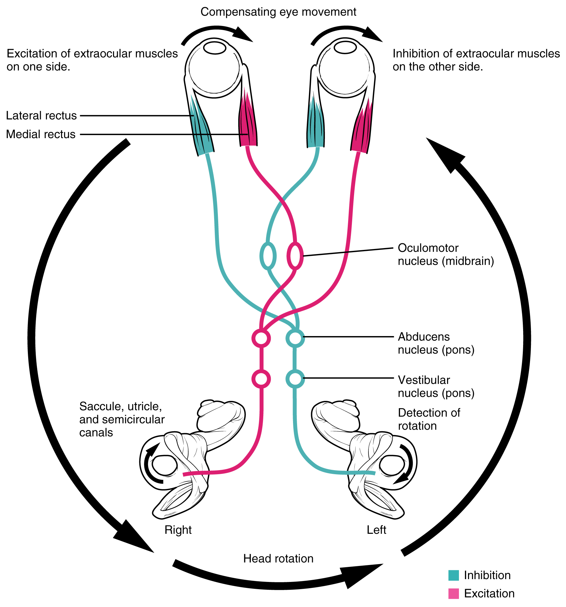
Neurons in the vestibular nuclei project their axons to targets in the brain stem. One target is the reticular formation, which influences respiratory and cardiovascular functions in relation to body movements. A second target of the axons of neurons in the vestibular nuclei is the spinal cord, which initiates the spinal reflexes involved with posture and balance. To assist the visual system, fibers of the vestibular nuclei project to the oculomotor, trochlear, and abducens nuclei to influence signals sent along the cranial nerves. These connections constitute the pathway of the vestibulo-ocular reflex (VOR), which compensates for head and body movement by stabilizing images on the retina (Figure 15.4.3). Finally, the vestibular nuclei project to the thalamus to join the proprioceptive pathway of the dorsal column system, allowing conscious perception of equilibrium.
Chapter Review
Like hearing, the ear detects head position, head movement, and when our bodies are in motion, all of which contribute to our sense of equilibrium. This information is related to the CNS by the vestibular branch of the vestibulocohlear nerve. Linear movements, like those experienced due to acceleration or tilting the head, are detected by the utricle and saccule based on the patterns of hair cell depolarization in response to the top-heavy otolithic membrane moving separately from the macula and bending the hair cells. Rotational movements move the head while semicircular canal fluid lags behind, deflecting the cupulae of the semicircular canals and activating hair cells. Three semicircular canals that can each be activated in two directions allows the vestibular system to detect the direction of most heads movements.
In the CNS, equilibrium information is used to maintain eye contact which moving, adjust posture and balance, and to adjust respiratory and cardiovascular function. Equilibrium stimuli are consciously perceived in the somatosensory cortex.
Glossary
- ampulla of the ear
- the structure at the base of a semicircular canal that contains the hair cells and cupula for transduction of rotational movement of the head
- cupula
- specialized structure within the base of a semicircular canal that bends the stereocilia of hair cells when the head rotates by way of the relative movement of the enclosed fluid
- equilibrium
- sense of balance that includes sensations of position and movement of the head
- macula
- enlargement at the base of a semicircular canal at which transduction of equilibrium stimuli takes place within the ampulla
- otolithic membrane
- gelatinous substance in the utricle and saccule of the inner ear that contains calcium carbonate crystals and into which the stereocilia of hair cells are embedded
- saccule
- structure of the inner ear responsible for transducing linear acceleration in the vertical plane
- semicircular canals
- structures within the inner ear responsible for transducing rotational movement information
- utricle
- structure of the inner ear responsible for transducing linear acceleration in the horizontal plane
- vestibular ganglion
- location of neuronal cell bodies that transmit equilibrium information along the eighth cranial nerve
- vestibular nuclei
- targets of the vestibular component of the eighth cranial nerve
- vestibulo-ocular reflex (VOR)
- reflex based on connections between the vestibular system and the cranial nerves of eye movements that ensures that images are stabilized on the retina as the head and body move
sense of balance that includes sensations of position and movement of the head
structure of the inner ear responsible for transducing linear acceleration in the horizontal plane
structure of the inner ear responsible for transducing linear acceleration in the vertical plane
structures within the inner ear responsible for transducing rotational movement information
location of neuronal cell bodies that transmit equilibrium information along the eighth cranial nerve
enlargement at the base of a semicircular canal at which transduction of equilibrium stimuli takes place within the ampulla
gelatinous substance in the utricle and saccule of the inner ear that contains calcium carbonate crystals and into which the stereocilia of hair cells are embedded
the structure at the base of a semicircular canal that contains the hair cells and cupula for transduction of rotational movement of the head
specialized structure within the base of a semicircular canal that bends the stereocilia of hair cells when the head rotates by way of the relative movement of the enclosed fluid
targets of the vestibular component of the eighth cranial nerve
reflex based on connections between the vestibular system and the cranial nerves of eye movements that ensures that images are stabilized on the retina as the head and body move
