11.7 Appendicular Muscles of the Pelvic Girdle and Lower Limbs
Learning Objectives
By the end of this section, you will be able to:
- Identify the appendicular muscles of the pelvic girdle and lower limbs as well as give their origins, insertions, actions and innervations
Appendicular Muscles of the Pelvic Girdle and Lower Limbs
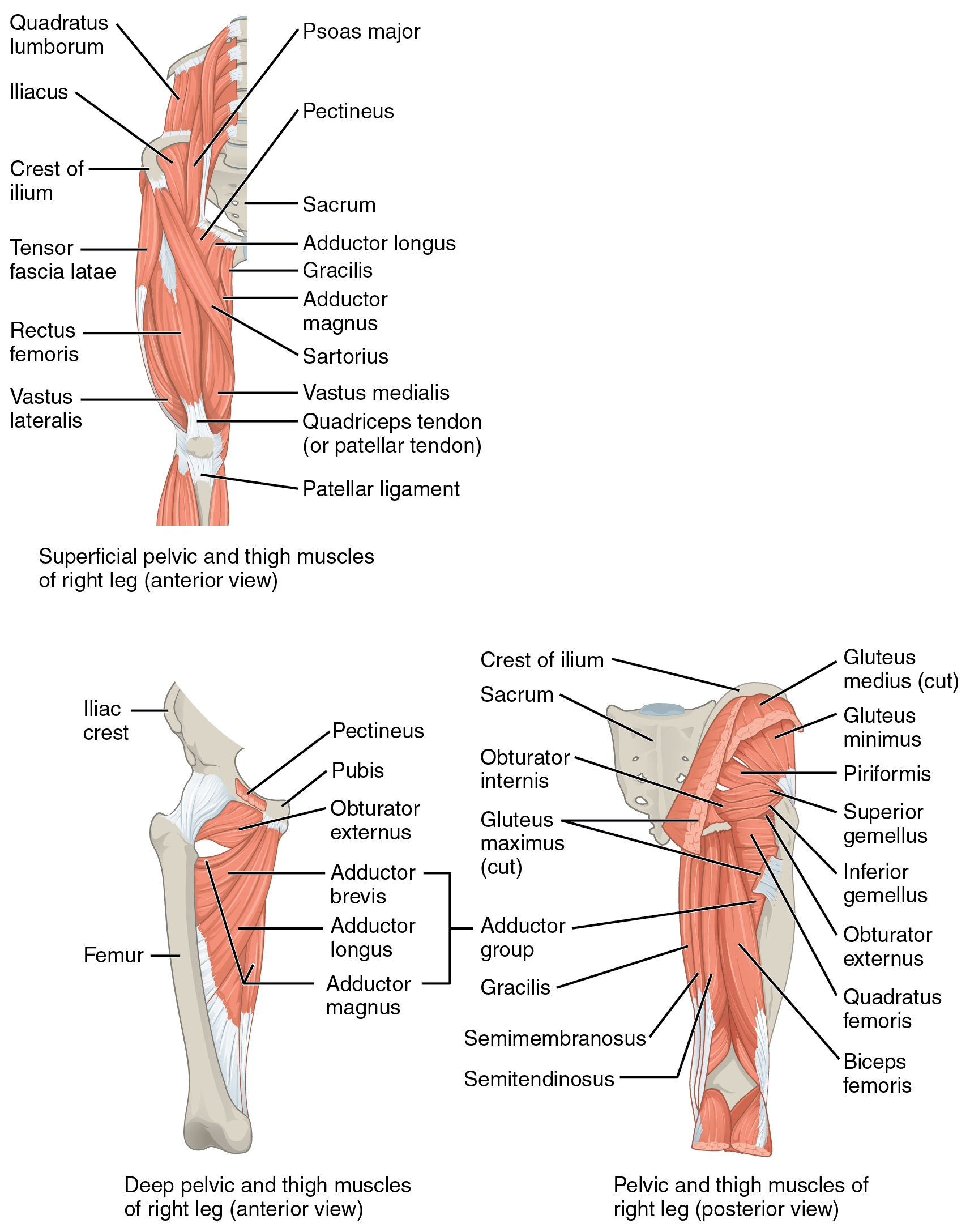
The appendicular muscles of the lower body position and stabilize the pelvic girdle, which serves as a foundation for the lower limbs. Comparatively, there is much more movement at the pectoral girdle than at the pelvic girdle. There is very little movement of the pelvic girdle because of its connection with the sacrum at the base of the axial skeleton and because the deep acetabulum provides a stable point of articulation with the head of the femur. The pelvic girdle’s lack of range of motion allows it to stabilize and support the body. The body’s center of gravity is in the area of the pelvis. If the center of gravity were not to remain fixed, standing up would be difficult. Therefore, what the leg muscles lack in range of motion and versatility, they make up for in size and power, facilitating the body’s stabilization, posture, and movement.
Gluteal Region Muscles That Move the Thigh
Most muscles that insert on the femur (the thigh bone) and move it, originate on the pelvic girdle. The major flexors of the hip are the psoas major and iliacus which make up the iliopsoas group. Some of the largest and most powerful muscles in the body are the gluteal muscles or gluteal group. The gluteus maximus, one of the major extensors of the thigh at the hip, is the largest; deep to the gluteus maximus is the gluteus medius, and deep to the gluteus medius is the gluteus minimus, the smallest of the trio (Figure 11.7.1 and Table 11.16a).
| Prime Mover | Movement | Target | Target Movement Direction | Origin | Insertion |
|---|---|---|---|---|---|
| Psoas major | Raises knee at hip, as if performing a knee attack; assists l rotators in twisting thigh (and lower leg) outward; assists with bending over, maintaining posture | Femur | Thigh: flexion and lateral rotation; torso: flexion | Lumbar vertebrae (L1-L5); thoracic vertebra (T12) | Lesser trochanter of femur |
| Iliacus | Raises knee at hip, as if performing a knee attack; assists l rotators in twisting thigh (and lower leg) outward; assists with bending over, maintaining posture | Femur | Thigh: flexion and lateral rotation; torso: flexion | Iliac fossa; iliac crest; lateral sacrum | Lesser trochanter of femur |
| Prime Mover | Movement | Target | Target Movement Direction | Origin | Insertion |
|---|---|---|---|---|---|
| Gluteus maximus | Lowers knee and move back, as when getting ready to kick a ball | Femur | Extension | Dorsal ilium; sacrum; coccyx | Gluteal tuberosity of femur; iliotibial tract |
| Gluteus medius | Open thighs, as when doing a split | Femur | Abduction | Lateral surface of ilium | Greater trochanter of femur |
| Gluteus minimus | Brings the thighs back together | Femur | Abduction | External surface of ilium | Greater trochanter of femur |
| Tensor facia lata | Assists with raising knee at hip and opening thighs; maintains posture by stabilizing the iliotibial track, which connects to the knee | Femur | Flexion; abduction | Anterior aspect of iliac crest; anterior superior iliac spine | Iliotibial tract |
| Prime Mover | Movement | Target | Target Movement Direction | Origin | Insertion |
|---|---|---|---|---|---|
| Piriformis | Twists thigh (and lower leg) outward; maintains posture by stabilizing hip joint | Femur | Lateral rotation | Anterolateral surface of sacrum | Greater trochanter of femur |
| Obturator internus | Twists thigh (and lower leg) outward; maintains posture by stabilizing hip joint | Femur | Lateral rotation | Inner surface of obturator membrane; greater sciatic notch; margins of obturator foramen | Greater trochanter in front of piriformis |
| Obturator externus | Twists thigh (and lower leg) outward; maintains posture by stabilizing hip joint | Femur | Lateral rotation | Outer surfaces of obturator membrane; pubic, and ischium; margins of obturator foramen | Trochanteric fossa of posterior femur |
| Superior gemellus | Twists thigh (and lower leg) outward; maintains posture by stabilizing hip joint | Femur | Lateral rotation | Ischial spine | Greater trochanter of femur |
| Inferior gemellus | Twists thigh (and lower leg) outward; maintains posture by stabilizing hip joint | Femur | Lateral rotation | Ischial tuberosity | Greater trochanter of femur |
| Quadratus femoris | Twists thigh (and lower leg) outward; maintains posture by stabilizing hip joint | Femur | Lateral rotation | Ischial tuberosity | Trochanteric crest of femur |
| Prime Mover | Movement | Target | Target Movement Direction | Origin | Insertion |
|---|---|---|---|---|---|
| Adductor longus | Brings the thighs back together; assists with raising the knee | Femur | Adduction; flexion | Pubis near pubic symphysis | Linea aspera |
| Adductor brevis | Brings the thighs back together; assists with raising the knee | Femur | Adduction; flexion | Body of pubis; inferior ramus of pubis | Linea aspera above adductor longus |
| Adductor magnus | Brings the thighs back together; assists with raising the knee and moving the thigh back | Femur | Adduction; flexion; extension | Ischial rami; pubic rami; ischial tuberosity | Linea aspera; adductor tubercle of femur |
| Pectineus | Opens thighs; assists with raising the knee and turning the thigh (and lower leg) inward | Femur | Adduction; flexion; medial rotation | Pectineal line of pubis | Lesser trochanter to linea aspera of posterior aspect of femur |
The tensor fascia latae is a thick, squarish muscle in the superior aspect of the lateral thigh. It acts as a synergist of the gluteus medius and iliopsoas in flexing and abducting the thigh. It also helps stabilize the lateral aspect of the knee by pulling on the iliotibial tract (band), making it taut. Deep to the gluteus maximus, the piriformis, obturator internus, obturator externus, superior gemellus, inferior gemellus, and quadratus femoris laterally rotate the thigh at the hip.
Deep fascia in the thigh separates it into medial, anterior, and posterior compartments. The muscles in the medial compartment of the thigh responsible for adducting the femur at the hip are the adductor group including the adductor longus, adductor brevis, and adductor magnus which all adduct and medially rotate the thigh. The adductor longus also flexes the thigh, whereas the adductor magnus extends it. Like the adductor longs, the pectineus adducts and flexes the femur at the hip. The pectineus is located in the femoral triangle, which is formed at the junction between the hip and the leg and includes the femoral nerve, the femoral artery, the femoral vein, and the deep inguinal lymph nodes. The strap-like gracilis adducts the thigh in addition to flexing the leg at the knee
Thigh Muscles That Move the Femur, Tibia, and Fibula
| Prime Mover | Movement | Target | Target Movement Direction | Origin | Insertion |
|---|---|---|---|---|---|
| Gracilis | Moves back of lower legs up toward buttocks, as when kneeling; assists in opening thighs | Femur; tibia/fibula | Tibia/fibula: flexion; thigh: adduction | Inferior ramus; body of pubis; ischial ramus | Medial surface of tibia |
| Prime Mover | Movement | Target | Target Movement Direction | Origin | Insertion |
|---|---|---|---|---|---|
| Rectus femoris | Moves lower leg out in front of body, as when kicking; assists in raising the knee | Femur; tibia/fibula | Tibia/fibula: extension; thigh: flexion | Anterior inferior iliac spine; superior margin of acetabulum | Patella; tibial tuberosity |
| Vastus lateralis | Moves lower leg out in front of body, as when kicking | Tibia/fibula | Extension | Greater trochanter; intertrochanteric line; linea aspera | Patella; tibial tuberosity |
| Vastus medialis | Moves lower leg out in front of body, as when kicking | Tibia/fibula | Extension | Linea aspera; intertrochanteric line | Patella; tibial tuberosity |
| Vastus intermedius | Moves lower leg out in front of body, as when kicking | Tibia/fibula | Extension | Proximal femur shaft | Patella; tibial tuberosity |
| Sartorius | Moves back of lower legs up toward buttocks, as when kneeling; assists in moving thigh diagonally upward and outward as when mounting a bike | Femur; tibia/fibula | Tibia: flexion; thigh: flexion, abduction, lateral rotation | Anterior inferior iliac spine | Medial aspect of proximal tibia |
| Prime Mover | Movement | Target | Target Movement Direction | Origin | Insertion |
|---|---|---|---|---|---|
| Biceps femoris | Moves back of lower legs up and back toward the buttocks, as when kneeling; moves thigh down and back; twists the thigh (and lower leg) outward | Femur; tibia/fibula | Tibia/fibula: flexion; thigh: extension, lateral rotation | Ischial tuberosity; linea aspera; distal femur | Head of fibula; lateral condyle of tibia |
| Semitendinosus | Moves back of lower legs up and back toward the buttocks, as when kneeling; moves thigh down and back; twists the thigh (and lower leg) inward | Femur; tibia/fibula | Tibia/fibula: flexion; thigh: extension, medial rotation | Ischial tuberosity | Upper tibial shaft |
| Semimembranosus | Moves back of lower legs up and back toward the buttocks, as when kneeling; moves thigh down and back; twists the thigh (and lower leg) inward | Femur; tibia/fibula | Tibia/fibula: flexion; thigh: extension, medial rotation | Ischial tuberosity | Medial condyle of tibia; lateral condyle of femur |
The muscles of the anterior compartment of the thigh flex the thigh and extend the leg. This compartment contains the quadriceps femoris group, which is comprised of four muscles that extend the leg and stabilize the knee. Within the compartment the rectus femoris is on the anterior aspect of the thigh, the vastus lateralis is on the lateral aspect of the thigh, the vastus medialis is on the medial aspect of the thigh, and the vastus intermedius is between the vastus lateralis and vastus medialis and deep to the rectus femoris. The tendon common to all four is the quadriceps tendon (patellar tendon), which inserts into the patella and continues below it as the patellar ligament. The patellar ligament attaches to the tibial tuberosity. In addition to the quadriceps femoris, the sartorius is a band-like muscle that extends from the anterior superior iliac spine to the medial side of the proximal tibia. This versatile muscle flexes the leg at the knee and flexes, abducts, and laterally rotates the thigh at the hip. This muscle allows us to sit cross-legged.
The posterior compartment of the thigh includes muscles that flex the leg and extend the thigh. The three long muscles on the back of the thigh are the hamstring group, which flexes the knee. These are the biceps femoris, semitendinosus, and semimembranosus. The tendons of these muscles form the upper border of the popliteal fossa, the diamond-shaped space at the back of the knee.
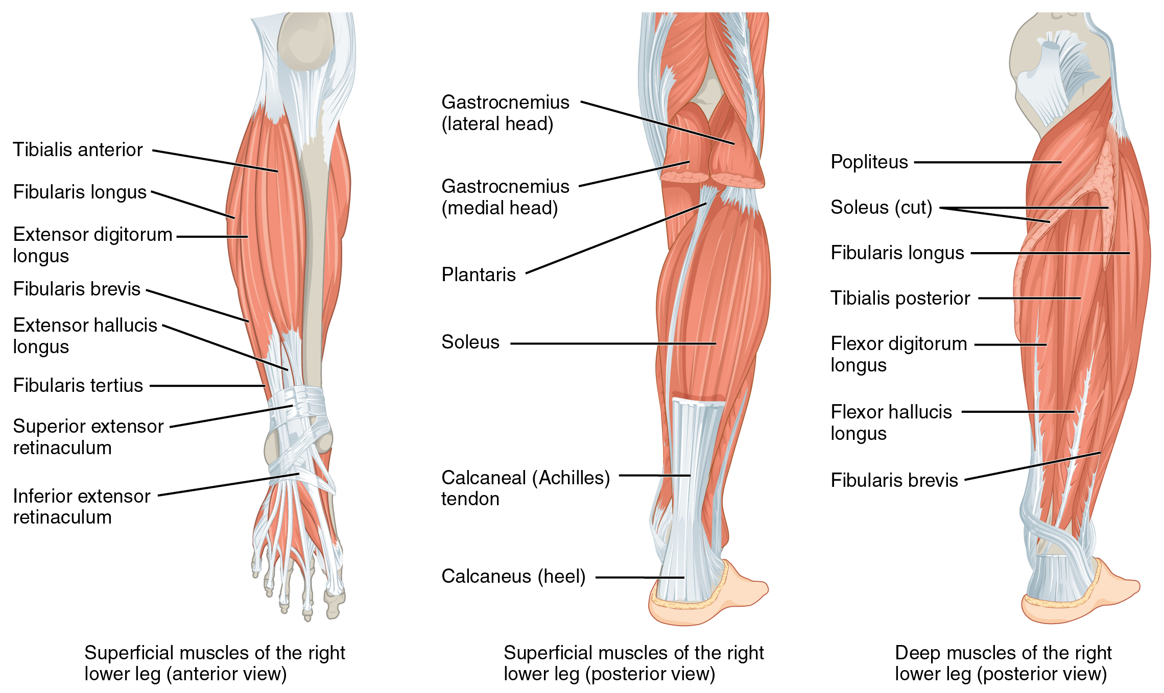
Muscles That Move the Feet and Toes
Similar to the thigh muscles, the muscles of the leg are divided by deep fascia into compartments, although the leg has three: anterior, lateral, and posterior.
| Prime Mover | Movement | Target | Target Movement Direction | Origin | Insertion |
|---|---|---|---|---|---|
| Tibialis anterior | Raises the sole of the foot off the ground, as when preparing to foot-tap; bends the inside of the foot upwards, as when catching your balance while falling laterally toward the opposite side as the balancing foot | Foot | Dorsiflexion; inversion | Lateral condyle and upper tibial shaft; interosseous membrane | Interior surface of medial cuneiform; First metatarsal bone |
| Extensor hallucis longus | Raises the sole of the foot off the ground, as when preparing to foot-tap; extends the big toe | Foot; big toe | Foot: dorsiflexion; big toe: extension | Anteromedial fibula shaft; interosseous membrane | Distal phalanx of big toe |
| Extensor digitorum longus | Raises the sole of the foot off the ground, as when preparing to foot-tap; extends toes | Foot; toes 2-5 | Foot: dorsiflexion; big toe: extension | Lateral condyle of tibia; proximal portion of fibula; interosseous membrane | Middle and distal phalanges of toes 2-5 |
| Prime Mover | Movement | Target | Target Movement Direction | Origin | Insertion |
|---|---|---|---|---|---|
| Fibularis longus | Lowers the sole of the foot to the ground, as when foot-tapping or jumping; bends the inside of the foot downward, as when catching your balance while falling laterally toward the same side as the balancing foot | Foot | Plantar flexion and eversion | Upper portion of lateral fibula | First metatarsal; medial cuneiform |
| Fibularis (peroneus) brevis | Lowers the sole of the foot to the ground, as when foot-tapping or jumping; bends the inside of the foot downward, as when catching your balance while falling laterally toward the same side as the balancing foot | Foot | Plantar flexion and eversion | Distal fibula shaft | Proximal end of fifth metatarsal |
| Prime Mover | Movement | Target | Target Movement Direction | Origin | Insertion |
|---|---|---|---|---|---|
| Gastrocnemius | Lowers the sole of the foot to the ground, as when foot-tapping or jumping; assists in moving the back of the lower legs up and back toward the buttocks | Foot; tibia/fibula | Foot: plantar flexion; tibia/fibula: flexion | Medial and lateral condyles of femur | Posterior calcaneus |
| Soleus | Lowers the sole of the foot to the ground, as when foot-tapping or jumping; maintains posture while walking | Foot | Plantar flexion | Superior tibia; fibula; interosseous membrane | Posterior calcaneus |
| Plantaris | Lowers the sole of the foot to the ground, as when foot-tapping or jumping; assists in moving the back of the lower legs up and back toward the buttocks | Foot; tibia/fibula | Foot: plantar flexion; tibia/fibula: flexion | Posterior femur above lateral condyle | Calcaneus or calcaneus tendon |
| Tibialis posterior | Lowers the sole of the foot to the ground, as when foot-tapping or jumping | Foot | Plantar flexion | Superior tibia and fibula; interosseous membrane | Several tarsal and metatarsals 2-4 |
| Prime Mover | Movement | Target | Target Movement Direction | Origin | Insertion |
|---|---|---|---|---|---|
| Popliteus | Moves the back of the lower legs up and back toward the buttocks; assists in the rotation of the leg at the knee and thigh | Tibia/fibula | Tibia/fibula: flexion thigh and lower leg: medial and lateral rotation | Lateral condyle of femur; lateral meniscus | Proximal tibia |
| Flexor digitorum longus | Lowers the sole of the foot to the ground, as when foot-tapping or jumping; bends the inside of the foot upward and flexes toes | Foot; toes 2-5 | Foot: plantar flexion and inversion toes: flexion | Posterior tibia | Distal phalanges of toes 2-5 |
| Flexor hallucis longus | Flexes the big toe | Big toe; foot | Big toe: flexion foot: plantar flexion | Midshaft of fibula; interosseous membrane | Distal phalanx of big toe |
The muscles in the anterior compartment of the leg all contribute to dorsiflexion: the tibialis anterior, a long and thick muscle on the lateral surface of the tibia, the extensor hallucis longus, deep under it, and the extensor digitorum longus, lateral to it. The fibularis tertius, a small muscle that originates on the anterior surface of the fibula, is associated with the extensor digitorum longus and sometimes fused to it, but is not present in all people. Thick bands of connective tissue called the superior extensor retinaculum (transverse ligament of the ankle) and the inferior extensor retinaculum, hold the tendons of these muscles in place during dorsiflexion.
The lateral compartment of the leg includes two muscles which contribute to eversion and plantar flexion: the fibularis longus (peroneus longus) and the fibularis brevis (peroneus brevis). The superficial muscles in the posterior compartment of the leg all insert onto the calcaneal tendon (Achilles tendon), a strong tendon that inserts into the calcaneal bone of the ankle, all contribute to plantar flexion. The muscles in this compartment are large and strong and keep humans upright. The most superficial and visible muscle of the calf is the gastrocnemius. Deep to the gastrocnemius is the wide, flat soleus. The plantaris runs obliquely between the two; some people may have two of these muscles, whereas no plantaris is observed in about seven percent of other cadaver dissections. The plantaris tendon is a desirable substitute for the fascia lata in hernia repair, tendon transplants, and repair of ligaments. There are four deep muscles in the posterior compartment of the leg as well: the popliteus, flexor digitorum longus, flexor hallucis longus, and tibialis posterior all contribute to plantar flexion or inversion of the foot.
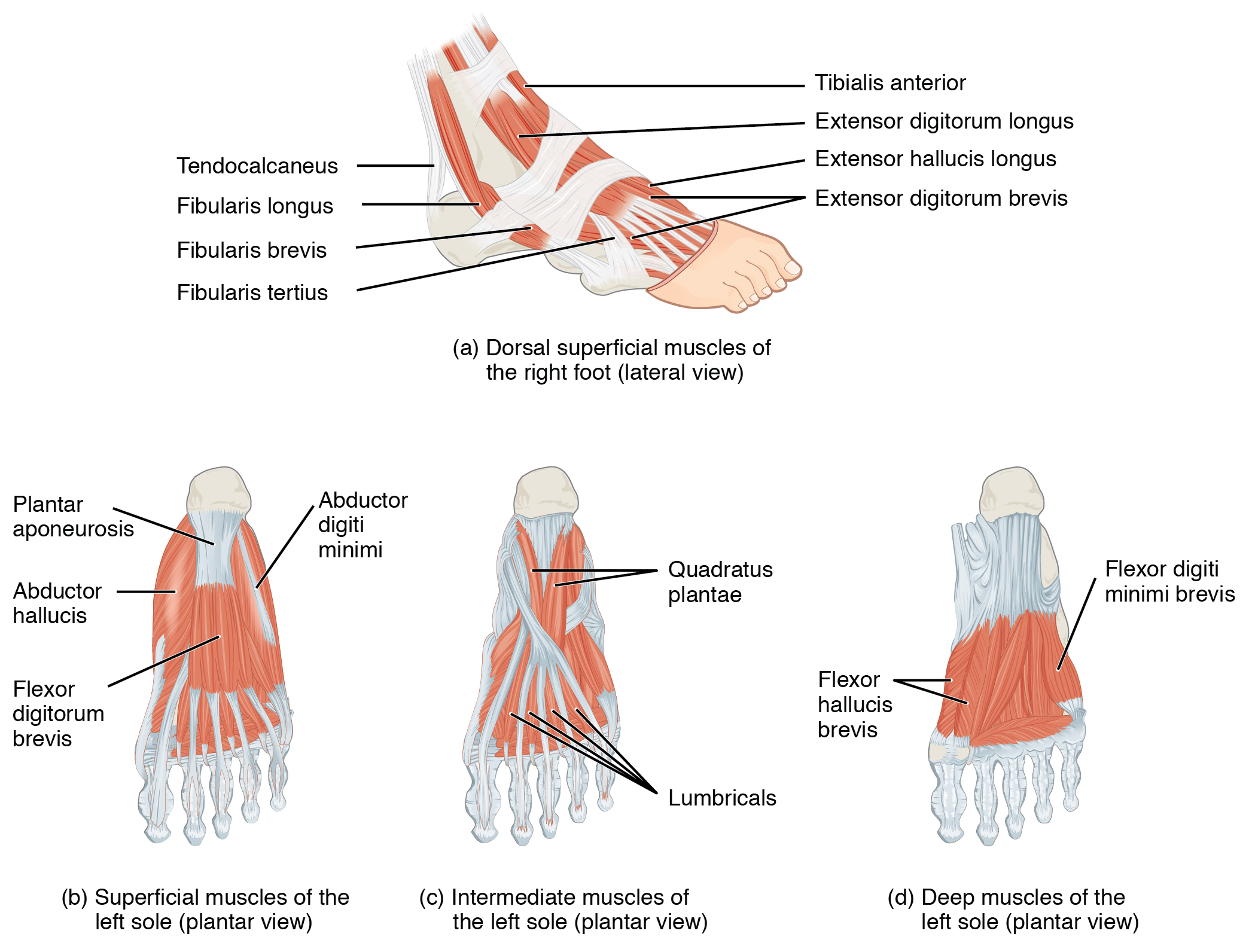
The foot also has intrinsic muscles, which originate and insert within it (similar to the intrinsic muscles of the hand). These muscles primarily provide support for the foot and its arch, and contribute to movements of the toes (Figure 11.7.3 and Table 11.19a). The principal support for the longitudinal arch of the foot is a deep fascia called plantar aponeurosis, which runs from the calcaneus bone to the toes (inflammation of this tissue is the cause of “plantar fasciitis,” which can affect runners. The intrinsic muscles of the foot include the extensor digitorum brevis on the dorsal aspect and a plantar group, which consists of four layers.
| Prime Mover | Movement | Target | Target Movement Direction | Origin | Insertion |
|---|---|---|---|---|---|
| Extensor digitorum brevis | Extends toes 2-5 | Toes 2-5 | Extension | Calcaneus; extensor retinaculum | Base of proximal phalanx of big toe; extensor expansions on toes 2-5 |
| Prime Mover | Movement | Target | Target Movement Direction | Origin | Insertion |
|---|---|---|---|---|---|
| Abductor hallucis | Abducts and flexes big toe | Big toe | Abduction; flexion | Calcaneal tuberosity; flexor retinaculum | Proximal phalanx of big toe |
| Flexor digitorum brevis | Flexes toes 2-4 | Middle toes | Flexion | Calcaneal tuberosity | Middle phalanx of toes 2-4 |
| Abductor digit minimi | Abducts and flexes small toe | Toe 5 | Abduction; flexion | Calcaneal tuberosity | Proximal phalanx of little toe |
| Prime Mover | Movement | Target | Target Movement Direction | Origin | Insertion |
|---|---|---|---|---|---|
| Quadratus plantae | Assists in flexing toes 2-5 | Toes 2-5 | Flexion | Medial and lateral sides of calcaneus | Tendon of flexor digitorum longus |
| Lumbricals | Extends toes 2-5 at the interphalangeal joints; flexes the small toes at the metatarsophalangeal joints | Toes 2-5 | Extension flexion | Tendons of flexor digitorum longus | Meidal side of proximal phalanx of toes 2-5 |
| Prime Mover | Movement | Target | Target Movement Direction | Origin | Insertion |
|---|---|---|---|---|---|
| Flexor hallucis | Flexes big toe | Big toe | Flexion | Lateral cuneiform; cuboid bones | Base of proximal phalanx of big toe |
| Adductor hallucis | Adducts and flexes big toe | Big toe | Abduction; flexion | Bases of metatarsals 2-4; fibularis longus tendon sheath; ligament across metatarsophalangeal joints | Base of proximal phalanx of big toe |
| Flexor digiti minimi brevis | Flexes small toe | Little toe | Flexion | base of metatarsal 5; tendon sheath of fibularis longus | Base of proximal phalanx of big toe |
| Prime Mover | Movement | Target | Target Movement Direction | Origin | Insertion |
|---|---|---|---|---|---|
| Dorsal interossei | Abducts and flexes middle toes at metatarsophalangeal joints; extends middle toes at interphalangeal joints | Middle toes | Abduction; flexion; extension | Side of metatarsals | Both sides of toe 2; for each other toe, extensor expansion over first phalanx on side opposite toe 2 |
| Plantar interossei | Abducts toes 3-5; flexes proximal phalanges and extends distal phalanges | Small toes | Abduction; flexion; extension | Side of each metatarsal that faces metatarsal 2 (absent from metatarsal 2) | Extensor expansion on first phalanx of each toe (except to 2) on side facing toe 2 |
Chapter Review
The pelvic girdle attaches the legs to the axial skeleton. The hip joint is where the pelvic girdle and the leg come together. The hip is joined to the pelvic girdle by many muscles. In the gluteal region, the psoas major and iliacus form the iliopsoas. The large and strong gluteus maximus, gluteus medius, and gluteus minimus extend and abduct the femur. Along with the gluteus maximus, the tensor fascia lata muscle forms the iliotibial tract. The lateral rotators of the femur at the hip are the piriformis, obturator internus, obturator externus, superior gemellus, inferior gemellus, and quadratus femoris. On the medial part of the thigh, the adductor longus, adductor brevis, and adductor magnus adduct the thigh and medially rotate it. The pectineus muscle adducts and flexes the femur at the hip.
The thigh muscles that move the femur, tibia, and fibula are divided into medial, anterior, and posterior compartments. The medial compartment includes the adductors, pectineus, and the gracilis. The anterior compartment comprises the quadriceps femoris, quadriceps tendon, patellar ligament, and the sartorius. The quadriceps femoris is made of four muscles: the rectus femoris, the vastus lateralis, the vastus medius, and the vastus intermedius, which together extend the knee. The posterior compartment of the thigh includes the hamstrings: the biceps femoris, semitendinosus, and the semimembranosus, which all flex the knee.
The muscles of the leg that move the foot and toes are divided into anterior, lateral, superficial- and deep-posterior compartments. The anterior compartment includes the tibialis anterior, the extensor hallucis longus, the extensor digitorum longus, and the fibularis (peroneus) tertius. The lateral compartment houses the fibularis (peroneus) longus and the fibularis (peroneus) brevis. The superficial posterior compartment has the gastrocnemius, soleus, and plantaris; and the deep posterior compartment has the popliteus, tibialis posterior, flexor digitorum longus, and flexor hallucis longus.
Review Questions
Critical Thinking Questions
Which muscles form the hamstrings? How do they function together?
Reveal
Which muscles form the quadriceps? How do they function together?
Reveal
Glossary
- adductor brevis
- muscle that adducts and medially rotates the thigh
- adductor longus
- muscle that adducts, medially rotates, and flexes the thigh
- adductor magnus
- muscle with an anterior fascicle that adducts, medially rotates and flexes the thigh, and a posterior fascicle that assists in thigh extension
- anterior compartment of the leg
- region that includes muscles that dorsiflex the foot
- anterior compartment of the thigh
- region that includes muscles that flex the thigh and extend the leg
- biceps femoris
- hamstring muscle
- calcaneal tendon
- (also, Achilles tendon) strong tendon that inserts into the calcaneal bone of the ankle
- extensor digitorum brevis
- muscle that extends the toes
- extensor digitorum longus
- muscle that is lateral to the tibialis anterior
- extensor hallucis longus
- muscle that is partly deep to the tibialis anterior and extensor digitorum longus
- femoral triangle
- region formed at the junction between the hip and the leg and includes the pectineus, femoral nerve, femoral artery, femoral vein, and deep inguinal lymph nodes
- fibularis brevis
- (also, peroneus brevis) muscle that plantar flexes the foot at the ankle and everts it at the intertarsal joints
- fibularis longus
- (also, peroneus longus) muscle that plantar flexes the foot at the ankle and everts it at the intertarsal joints
- fibularis tertius
- small muscle that is associated with the extensor digitorum longus
- flexor digitorum longus
- muscle that flexes the four small toes
- flexor hallucis longus
- muscle that flexes the big toe
- gastrocnemius
- most superficial muscle of the calf
- gluteal group
- muscle group that extends, flexes, rotates, adducts, and abducts the femur
- gluteus maximus
- largest of the gluteus muscles that extends the femur
- gluteus medius
- muscle deep to the gluteus maximus that abducts the femur at the hip
- gluteus minimus
- smallest of the gluteal muscles and deep to the gluteus medius
- gracilis
- muscle that adducts the thigh and flexes the leg at the knee
- hamstring group
- three long muscles on the back of the leg
- iliacus
- muscle that, along with the psoas major, makes up the iliopsoas
- iliopsoas group
- muscle group consisting of iliacus and psoas major muscles, that flexes the thigh at the hip, rotates it laterally, and flexes the trunk of the body onto the hip
- iliotibial tract
- muscle that inserts onto the tibia; made up of the gluteus maximus and connective tissues of the tensor fasciae latae
- inferior extensor retinaculum
- cruciate ligament of the ankle
- inferior gemellus
- muscle deep to the gluteus maximus on the lateral surface of the thigh that laterally rotates the femur at the hip
- lateral compartment of the leg
- region that includes the fibularis (peroneus) longus and the fibularis (peroneus) brevis and their associated blood vessels and nerves
- medial compartment of the thigh
- a region that includes the adductor longus, adductor brevis, adductor magnus, pectineus, gracilis, and their associated blood vessels and nerves
- obturator externus
- muscle deep to the gluteus maximus on the lateral surface of the thigh that laterally rotates the femur at the hip
- obturator internus
- muscle deep to the gluteus maximus on the lateral surface of the thigh that laterally rotates the femur at the hip
- patellar ligament
- ligament spanning from the patella to the anterior tibia; extension of the quadriceps tendon which serves as the distal attachment for the muscles of the quadriceps femoris
- pectineus
- muscle that abducts and flexes the femur at the hip
- pelvic girdle
- (also, hip girdle) consists of a single hip bone, which attaches a lower limb to the axial skeleton at the sacrum
- piriformis
- muscle deep to the gluteus maximus on the lateral surface of the thigh that laterally rotates the femur at the hip
- plantar aponeurosis
- muscle that supports the longitudinal arch of the foot
- plantar group
- four-layered group of intrinsic foot muscles
- plantaris
- muscle that runs obliquely between the gastrocnemius and the soleus
- popliteal fossa
- diamond-shaped space at the back of the knee
- popliteus
- muscle that flexes the leg at the knee and creates the floor of the popliteal fossa
- posterior compartment of the leg
- region that includes the superficial gastrocnemius, soleus, and plantaris, and the deep popliteus, flexor digitorum longus, flexor hallucis longus, and tibialis posterior
- posterior compartment of the thigh
- region that includes muscles that flex the leg and extend the thigh
- psoas major
- muscle that, along with the iliacus, makes up the iliopsoas
- quadratus femoris
- muscle deep to the gluteus maximus on the lateral surface of the thigh that laterally rotates the femur at the hip
- quadriceps femoris group
- four muscles, that extend and stabilize the knee
- quadriceps tendon
- (also, patellar tendon) tendon common to all four quadriceps muscles, inserts into the patella
- rectus femoris
- quadricep muscle on the anterior aspect of the thigh
- sartorius
- band-like muscle that flexes, abducts, and laterally rotates the leg at the hip
- semimembranosus
- hamstring muscle
- semitendinosus
- hamstring muscle
- soleus
- wide, flat muscle deep to the gastrocnemius
- superior extensor retinaculum
- transverse ligament of the ankle
- superior gemellus
- muscle deep to the gluteus maximus on the lateral surface of the thigh that laterally rotates the femur at the hip
- tensor fascia latae
- muscle that flexes and abducts the thigh
- tibialis anterior
- muscle located on the lateral surface of the tibia
- tibialis posterior
- muscle that plantar flexes and inverts the foot
- vastus intermedius
- quadricep muscle that is between the vastus lateralis and vastus medialis and is deep to the rectus femoris
- vastus lateralis
- quadricep muscle on the lateral aspect of the thigh
- vastus medialis
- quadricep muscle on the medial aspect of the thigh
(also, hip girdle) consists of a single hip bone, which attaches a lower limb to the axial skeleton at the sacrum
muscle that, along with the iliacus, makes up the iliopsoas
muscle that, along with the psoas major, makes up the iliopsoas
muscle group consisting of iliacus and psoas major muscles, that flexes the thigh at the hip, rotates it laterally, and flexes the trunk of the body onto the hip
muscle group that extends, flexes, rotates, adducts, and abducts the femur
largest of the gluteus muscles that extends the femur
muscle deep to the gluteus maximus that abducts the femur at the hip
smallest of the gluteal muscles and deep to the gluteus medius
muscle that flexes and abducts the thigh
muscle that inserts onto the tibia; made up of the gluteus maximus and connective tissues of the tensor fasciae latae
muscle deep to the gluteus maximus on the lateral surface of the thigh that laterally rotates the femur at the hip
muscle deep to the gluteus maximus on the lateral surface of the thigh that laterally rotates the femur at the hip
muscle deep to the gluteus maximus on the lateral surface of the thigh that laterally rotates the femur at the hip
muscle deep to the gluteus maximus on the lateral surface of the thigh that laterally rotates the femur at the hip
muscle deep to the gluteus maximus on the lateral surface of the thigh that laterally rotates the femur at the hip
muscle deep to the gluteus maximus on the lateral surface of the thigh that laterally rotates the femur at the hip
a region that includes the adductor longus, adductor brevis, adductor magnus, pectineus, gracilis, and their associated blood vessels and nerves
muscle that adducts, medially rotates, and flexes the thigh
muscle that adducts and medially rotates the thigh
muscle with an anterior fascicle that adducts, medially rotates and flexes the thigh, and a posterior fascicle that assists in thigh extension
muscle that abducts and flexes the femur at the hip
region formed at the junction between the hip and the leg and includes the pectineus, femoral nerve, femoral artery, femoral vein, and deep inguinal lymph nodes
muscle that adducts the thigh and flexes the leg at the knee
region that includes muscles that flex the thigh and extend the leg
four muscles, that extend and stabilize the knee
quadricep muscle on the anterior aspect of the thigh
quadricep muscle on the lateral aspect of the thigh
quadricep muscle on the medial aspect of the thigh
quadricep muscle that is between the vastus lateralis and vastus medialis and is deep to the rectus femoris
(also, patellar tendon) tendon common to all four quadriceps muscles, inserts into the patella
ligament spanning from the patella to the anterior tibia; extension of the quadriceps tendon which serves as the distal attachment for the muscles of the quadriceps femoris
band-like muscle that flexes, abducts, and laterally rotates the leg at the hip
region that includes muscles that flex the leg and extend the thigh
three long muscles on the back of the leg
hamstring muscle
hamstring muscle
hamstring muscle
diamond-shaped space at the back of the knee
region that includes muscles that dorsiflex the foot
muscle located on the lateral surface of the tibia
muscle that is partly deep to the tibialis anterior and extensor digitorum longus
muscle that is lateral to the tibialis anterior
small muscle that is associated with the extensor digitorum longus
transverse ligament of the ankle
cruciate ligament of the ankle
region that includes the fibularis (peroneus) longus and the fibularis (peroneus) brevis and their associated blood vessels and nerves
(also, peroneus longus) muscle that plantar flexes the foot at the ankle and everts it at the intertarsal joints
(also, peroneus brevis) muscle that plantar flexes the foot at the ankle and everts it at the intertarsal joints
region that includes the superficial gastrocnemius, soleus, and plantaris, and the deep popliteus, flexor digitorum longus, flexor hallucis longus, and tibialis posterior
(also, Achilles tendon) strong tendon that inserts into the calcaneal bone of the ankle
most superficial muscle of the calf
wide, flat muscle deep to the gastrocnemius
muscle that runs obliquely between the gastrocnemius and the soleus
muscle that flexes the leg at the knee and creates the floor of the popliteal fossa
muscle that flexes the four small toes
muscle that flexes the big toe
muscle that plantar flexes and inverts the foot
muscle that supports the longitudinal arch of the foot
muscle that extends the toes
four-layered group of intrinsic foot muscles
